. 2021 Jan 28;13(2):193.
doi: 10.3390/v13020193.

Investigating the Diversity and Host Range of Novel Parvoviruses from North American Ducks Using Epidemiology, Phylogenetics, Genome Structure, and Codon Usage Analysis

Affiliations

Investigating the Diversity and Host Range of Novel Parvoviruses from North American Ducks Using Epidemiology, Phylogenetics, Genome Structure, and Codon Usage Analysis

Marta Canuti et al. Viruses. .

Abstract

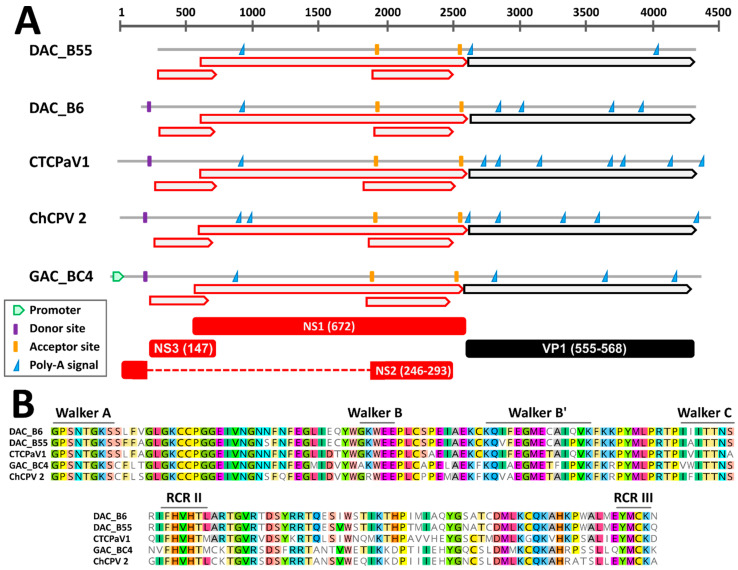
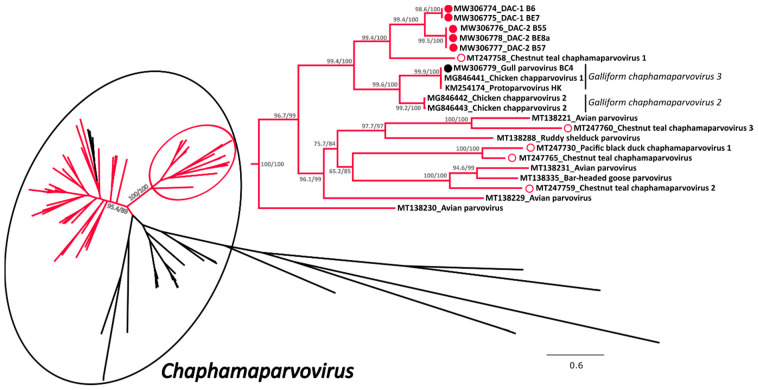
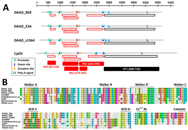
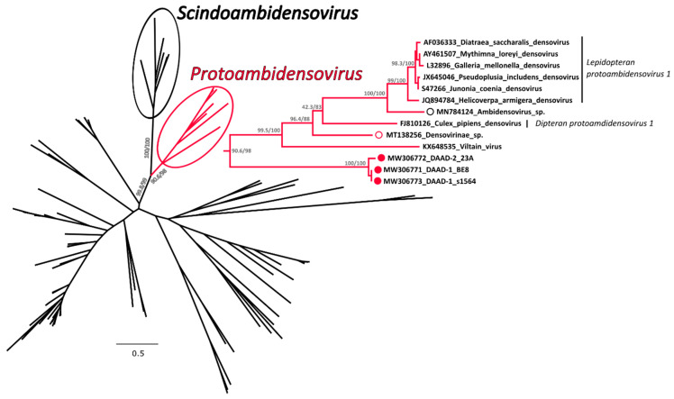
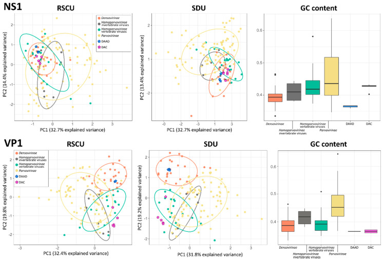
Parvoviruses are small single-stranded DNA viruses that can infect both vertebrates and invertebrates. We report here the full characterization of novel viruses we identified in ducks, including two viral species within the subfamily Hamaparvovirinae (duck-associated chapparvovirus, DAC) and a novel species within the subfamily Densovirinae (duck-associated ambidensovirus, DAAD). Overall, 5.7% and 21.1% of the 123 screened ducks (American black ducks, mallards, northern pintail) were positive for DAC and DAAD, respectively, and both viruses were more frequently detected in autumn than in winter. Genome organization and predicted transcription profiles of DAC and DAAD were similar to viruses of the genera Chaphamaparvovirus and Protoambidensovirus, respectively. Their association to these genera was also demonstrated by subfamily-wide phylogenetic and distance analyses of non-structural protein NS1 sequences. While DACs were included in a highly supported clade of avian viruses, no definitive conclusions could be drawn about the host type of DAAD because it was phylogenetically close to viruses found in vertebrates and invertebrates and analyses of codon usage bias and nucleotide frequencies of viruses within the family Parvoviridae showed no clear host-based viral segregation. This study highlights the high parvoviral diversity in the avian reservoir with many avian-associated parvoviruses likely yet to be discovered.

Keywords: avian viruses; chaphamaparvovirus; codon usage; densovirus; dinucleotide frequencies; insect viruses; parvovirus; virus discovery.


Conflict of interest statement

The authors declare no conflict of interest.

Figures

Figure 1
Figure 1
Genomic features of the novel duck-associated chapparvoviruses (DAC) compared to closely related viruses. (A) Schematic genome representations with in silico predicted open reading frames (ORFs, red for non-structural proteins (NS) and black for viral proteins (VP)), splicing donor and acceptor sites, poly-adenylation signals, and promoter (as indicated by the legend). Hypothetical proteins (with respective protein sizes) generated after mRNA splicing are shown at the bottom. (B) Conserved rolling circle replication (RCR) and Walker motifs typical of parvoviral SF3 helicases. CTCPaV1: Chestnut teal chaphamaparvovirus 1, ChCPV 2: chicken chapparvovirus 2, GAC: gull-associated chapparvovirus.
Figure 2
Figure 2
Phylogenetic analysis of the duck associated chapparvoviruses (DAC) within the Hamaparvovirinae. The phylogenetic tree based on 101 full NS1 protein sequences was built with the maximum-likelihood method based on the General matrix (LG) + F + R6 model with IQ-Tree [31]. The outcomes of the SH-aLRT and bootstrap test are shown for the main nodes. The branches of the unrooted tree are color-coded based on the host in which viruses have been identified and red represents avian hosts, while black includes all other vertebrate and invertebrate hosts. The black circle indicates viruses within the genus Chaphamaparvovirus while the red circle shows the clade containing the viruses studied here, which is also shown enlarged on the right side. The viruses identified in this study are labelled with a colored full circle (red for those found in ducks and black for the one found in a gull), while viruses found by others in ducks (genus Anas) are indicated by an empty red circle. Species designations, when available, are indicated on the right.
Figure 3
Figure 3
Genomic features of the novel duck-associated ambidensoviruses (DAAD) compared to that of Culex pipiens densovirus (CpDV). (A) Schematic genome representations with in silico predicted ORFs (red for NS and black for VP proteins), splicing donor and acceptor sites, poly-adenylation signals, and promoter (as indicated by the legend). Hypothetical proteins (with respective protein sizes) generated after mRNA splicing are shown at the bottom. (B) Conserved rolling circle replication (RCR) and Walker motifs typical of parvoviral SF3 helicases and phospholipase A2 (Ca++ BL: calcium binding loop; Catalytic: catalytic site) motifs typical of parvoviral VP1. GmDNV: Galleria mellonella densovirus.
Figure 4
Figure 4
Phylogenetic analysis of the duck associated ambidensovirus (DAAD) within the Densovirinae. The phylogenetic tree based on 117 full NS1 protein sequences was built with the maximum-likelihood method based on the General matrix (LG) + F + R6 model with IQ-Tree [31]. The outcomes of the SH-aLRT and bootstrap test are shown for main nodes. The branches of the unrooted tree are color-coded based on the taxonomy and red represents putative members of the genus Protoambidensovirus (enclosed in a red circle and shown in extenso on the right), while black includes all other viruses. The black circle indicates viruses within the genus Scindoambidensovirus. The viruses identified in this study are labelled with a full red circle, while viruses found by others in vertebrates are indicated by an empty red and black circle for avian and mammal studies, respectively. Species designations, when available, are indicated on the right.
Figure 5
Figure 5
Compositional analyses of viruses within the Parvoviridae. Principal component analysis (PCA) plots based on relative synonymous codon usage (RSCU) and synonymous dinucleotide usage (SDU) values are shown on the left and in the center, respectively; ellipses are drawn around groups as indicated in the legend with a normal probability size of 70% and axes represent dimensions explaining the largest variances. Box-and-whisker plots based on GC contents in each group are showed on the right; upper and lower whiskers indicate the highest and lowest values within 1.5 times the interquartile range, while boxes indicate the interquartile range itself with average values displayed as thick lines. Dots indicate potential outliers.

References

    1. Cotmore S.F., Agbandje-McKenna M., Canuti M., Chiorini J.A., Eis-Hubinger A.-M., Hughes J., Mietzsch M., Modha S., Ogliastro M., Pénzes J.J., et al. ICTV virus taxonomy profile: Parvoviridae. J. Gen. Virol. 2019;100:367–368. doi: 10.1099/jgv.0.001212. - DOI - PMC - PubMed
    1. Pénzes J.J., Söderlund-Venermo M., Canuti M., Eis-Hübinger A.M., Hughes J., Cotmore S.F., Harrach B. Reorganizing the family Parvoviridae: A revised taxonomy independent of the canonical approach based on host association. Arch. Virol. 2020;165:2133–2146. doi: 10.1007/s00705-020-04632-4. - DOI - PubMed
    1. Pénzes J.J., Canuti M., Söderlund-Venermo M., Eis-Huebinger A.M., Ogliastro M., Harrach B. ICTV Taxonomy Proposal 2020. International Committee on Taxonomy of Viruses (ICTV); 2020. Create three new genera and 19 new species (Piccovirales: Parvoviridae)
    1. Mietzsch M., Pénzes J.J., Agbandje-McKenna M. Twenty-five years of structural parvovirology. Viruses. 2019;11:362. doi: 10.3390/v11040362. - DOI - PMC - PubMed
    1. Cotmore S.F., Tattersall P. Parvoviruses: Small does not mean simple. Ann. Rev. Virol. 2014;1:517–537. doi: 10.1146/annurev-virology-031413-085444. - DOI - PubMed

Publication types

LinkOut - more resources