. 2021 Jan 28;13(2):196.
doi: 10.3390/v13020196.

Antiviral Activity of the G-Quadruplex Ligand TMPyP4 against Herpes Simplex Virus-1

Affiliations

Antiviral Activity of the G-Quadruplex Ligand TMPyP4 against Herpes Simplex Virus-1

Sara Artusi et al. Viruses. .

Abstract

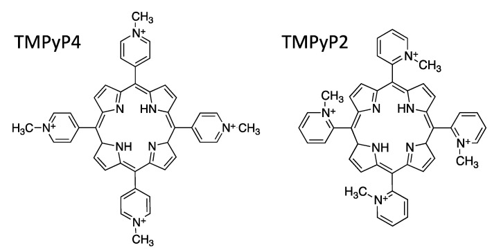
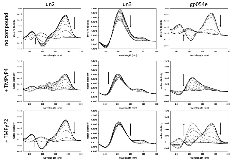
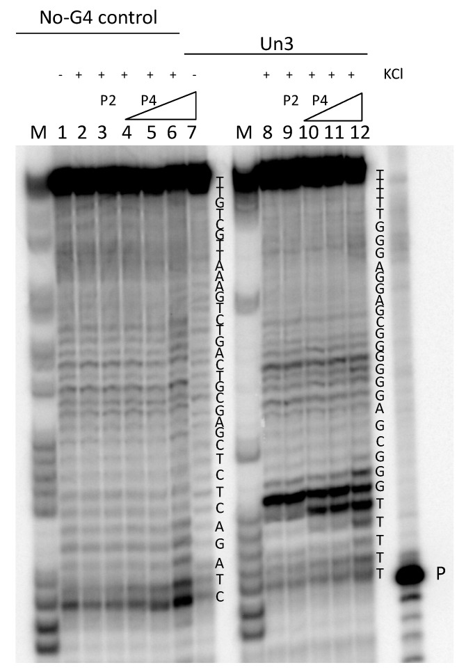
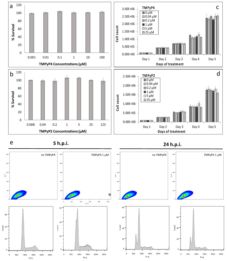
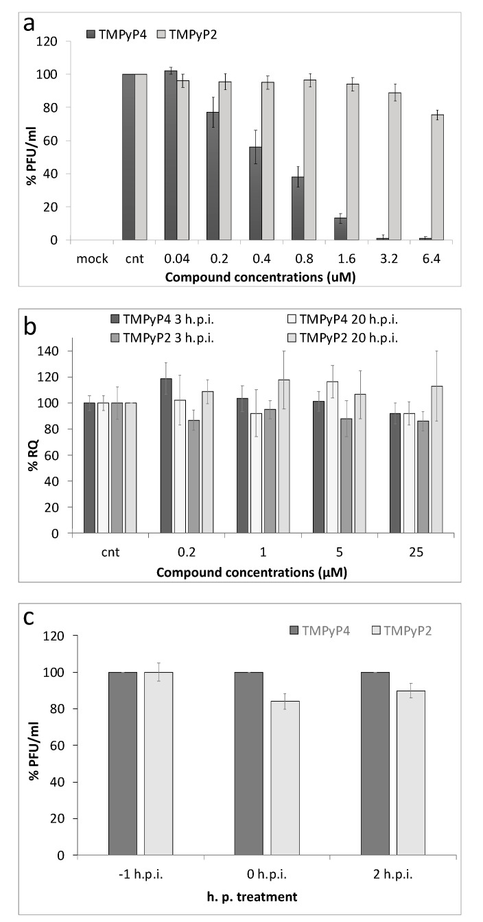
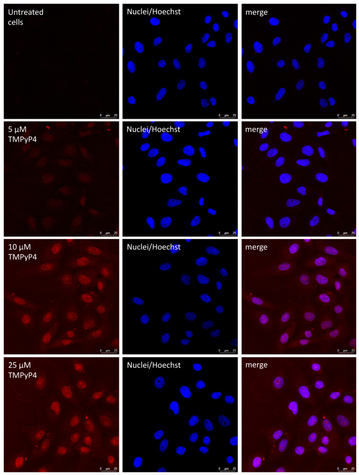
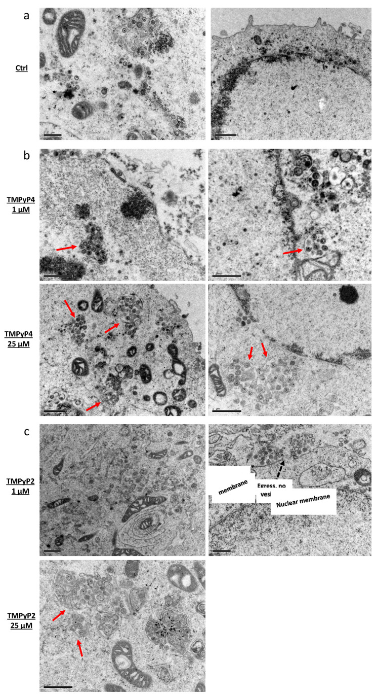
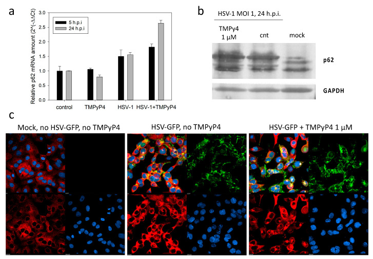
The herpes simplex virus 1 (HSV-1) genome is extremely rich in guanine tracts that fold into G-quadruplexes (G4s), nucleic acid secondary structures implicated in key biological functions. Viral G4s were visualized in HSV-1 infected cells, with massive virus cycle-dependent G4-formation peaking during viral DNA replication. Small molecules that specifically interact with G4s have been shown to inhibit HSV-1 DNA replication. We here investigated the antiviral activity of TMPyP4, a porphyrin known to interact with G4s. The analogue TMPyP2, with lower G4 affinity, was used as control. We showed by biophysical analysis that TMPyP4 interacts with HSV-1 G4s, and inhibits polymerase progression in vitro; in infected cells, it displayed good antiviral activity which, however, was independent of inhibition of virus DNA replication or entry. At low TMPyP4 concentration, the virus released by the cells was almost null, while inside the cell virus amounts were at control levels. TEM analysis showed that virus particles were trapped inside cytoplasmatic vesicles, which could not be ascribed to autophagy, as proven by RT-qPCR, western blot, and immunofluorescence analysis. Our data indicate a unique mechanism of action of TMPyP4 against HSV-1, and suggest the unprecedented involvement of currently unknown G4s in viral or antiviral cellular defense pathways.

Keywords: G-quadruplex; G-quadruplex ligands; HSV-1; TMPyP2; TMPyP4; antiviral activity.


Conflict of interest statement

The authors declare no conflict of interest. The funders had no role in the design of the study; in the collection, analyses, or interpretation of data; in the writing of the manuscript, or in the decision to publish the results.

Figures

Figure 1
Figure 1
Chemical structures of the cationic porphyrin 5,10,15,20-tetra-(N-methyl-4-pyridyl)porphyrin (TMPyP4) and the positional isomeric cationic porphyrin 5,10,15,20-tetra-(N-methyl-2-pyridyl)porphyrin (TMPyP2). TMPyP4 is a G4-ligand and is the compound under study; TMPyP2 has a lower affinity for G4s, and was used here as negative control [29,30,31].
Figure 2
Figure 2
Thermal unfolding spectra of three HSV-1 G4-forming sequences, un2, un3, and gp054e (4 μM), alone or in the presence of TMPyP4 or TMPyP2 (16 μM). Arrows in circular dichroism (CD) spectra indicate direction of the spectra upon temperature increase/DNA denaturation. Spectra were recorded over a temperature range of 20–90 °C.
Figure 3
Figure 3
Representative Taq polymerase stop gel. Oligonucleotides were folded in the presence or absence of K+. K+-treated samples were further incubated with increasing concentrations (125 nM, 250 nM, and 500 nM) of TMPyP4 (P4) or the control compound TMPyP2 (P2) (500 nM). Oligonucleotides were used as templates in a Taq polymerase reaction at 60 °C. The G4-forming oligonucleotide un3, and an oligonucleotide that does not form G4 (no-G4 control), were employed. Oligonucleotide sequences are indicated on the right of each set of samples. P indicates the band of the labeled primer. M is a marker lane obtained with the Maxam and Gilbert sequencing protocol.
Figure 4
Figure 4
Effect of TMPyP4 on uninfected Vero cells. (a) Percentage of cell survival determined by MTT following treatment with TMPyP4 (a) or TMPyP2 (b). For cell count, cells were treated with either TMPyP4 (c) or TMPyP2 (d), and counted every 24 h, for a total of five days. Two replicates per analysis were performed. (e) Flow cytometry analysis of Vero cells in the presence of TMPyP4. Vero cells gated for granularity (SSC) and size (FSC) and relative cell cycle histograms. Analysis was performed at 5 h and 24 h post treatment, as indicated, in the absence and presence of TMPyP4 1 μM.
Figure 5
Figure 5
Activity of TMPyP4 in HSV-1 infected cells. (a) Antiviral activity of TMPyP4 determined through plaque assay: HSV-1 infected cells (multiplicity of infection (MOI) 1 plaque forming units (PFU)/cell) were treated with increasing concentrations (0.04 µM–6.4 µM) of TMPyP4 or TMPyP2, used as a negative control; supernatants were collected 24 h.p.i., and the number of PFU was determined; (b) Levels of intracellular HSV-1 DNA, extracted from infected Vero cells treated with the test compounds. Quantification of intracellular DNA amounts was obtained from infected cells at 3 h and 20 h.p.i., treated with increasing concentration of TMPyP4 and TMPyP2. RQ is relative quantities; (c) Effect of TMPyP4 and TMPyP2 (1 µM) on virus entry into the cells. Cells were treated at various time points (−1, 0, and 2 h) relative to infection with HSV-1 strain F at a MOI of 1. One h after infection, cells were washed and maintained in culture medium supplemented with the drug. At 30 h.p.i. supernatants were collected and titrated by plaque assay. Virus yields are given as % relative to the 0 h.p.i. TMPyP4-treated sample.
Figure 6
Figure 6
Confocal microscopy images of Vero cells untreated or treated with 5, 10, and 25 μM TMPyP4 for 20 h. Cell nuclei were stained with Hoechst. The images show that the cationic porphyrin mainly localized in the cell nucleus.
Figure 7
Figure 7
Levels of infective virions were determined through plaque assay, both in supernatants and in cell lysates at 24 h.p.i., upon treatment with TMPyP4 (a), and with TMPyP2 (b) used as negative control.
Figure 8
Figure 8
Transmission electron microscopy analysis of HSV-1 infected cells at a MOI of 1, in the absence or presence of TMPyP4 or TMPyP2. Panels display: (a) non-treated infected Vero cells; (b) infected cells treated with TMPyP4 at 1 µM (top) or 25 µM (bottom); (c) infected cells treated with TMPyP2 at 1 µM (top) or 25 µM (bottom). In the cytoplasm of infected/treated cells HSV-1-containing vesicles are highlighted with solid red arrows. Dashed black arrows indicate viral egress from the cytoplasmatic membrane. Black bars at the bottom left corner of each figure indicate the image scale.
Figure 9
Figure 9
Expression of p62 upon treatment with TMPyP4. (a) p62 mRNA levels were analyzed by qRT-PCR. Uninfected and untreated cells were used as controls. Two replicates were performed. Standard deviation is shown. (b) p62 protein levels were determined through western blotting from lysates of Vero cells infected with HSV-1 at a of MOI of 1, in the absence or presence of TMPyP4 (1 μM). From right to left, lanes represent non-infected/non-treated Vero cells only, infected/non-treated Vero cells, infected/TMPyP4-treated cells. The housekeeping GAPDH (bottom lane) was used as a loading control. Three replicates were performed. (c) For immunofluorescence studies, Vero cells were infected with a GFP-VP16 HSV-1 (HSV-1 v41) in the absence (central panel) or presence of the G4-ligand TMPyP4 (1 μM, right panel). Mock Vero cells are shown on the left panel. Following fixation, permeabilization, and blocking, cells were probed with anti-p62 primary antibody and Alexa-546 secondary antibody. The green signal corresponds to the viral protein VP16-GFP, the red signal indicates p62, the blue signal indicates cell nuclei. Three replicates were performed.
Figure 10
Figure 10
RT-qPCR analysis of mRNA of the viral genes that express UL36, UL30, ICP22, and ICP34.5. Relative viral mRNA expression upon treatment with TMPyP4 (1 µM) is reported relative to the same sample without treatment (value of 1). Thus, samples that have relative mRNA expression higher and lower than 1 have higher and lower mRNA expression relative to their untreated control. All experiments were performed at least twice. Standard deviation is shown in all analyses.

References

    1. Looker K.J., Magaret A.S., May M.T., Turner K.M.E., Vickerman P., Gottlieb S.L., Newman L.M. Global and Regional Estimates of Prevalent and Incident Herpes Simplex Virus Type 1 Infections in 2012. PLoS ONE. 2015;10:e0140765. doi: 10.1371/journal.pone.0140765. - DOI - PMC - PubMed
    1. Frangoul H., Wills M., Crossno C., Engel M., Domm J. Acyclovir-resistant herpes simplex virus pneumonia post-unrelated stem cell transplantation: A word of caution. Pediatric Transplant. 2007;11:942–944. doi: 10.1111/j.1399-3046.2007.00822.x. - DOI - PubMed
    1. Roizman B., Whitley R.J. An Inquiry into the Molecular Basis of HSV Latency and Reactivation. Annu. Rev. Microbiol. 2013;67:355–374. doi: 10.1146/annurev-micro-092412-155654. - DOI - PubMed
    1. Hodge R.A.V., Field H.J. Chapter One—Antiviral Agents for Herpes Simplex Virus. In: De Clercq E., editor. Advances in Pharmacology. Academic Press; Cambridge, MA, USA: 2013. pp. 1–38. - DOI - PubMed
    1. Bacon T.H., Levin M.J., Leary J.J., Sarisky R.T., Sutton D. Herpes Simplex Virus Resistance to Acyclovir and Penciclovir after Two Decades of Antiviral Therapy. Clin. Microbiol. Rev. 2003;16:114–128. doi: 10.1128/CMR.16.1.114-128.2003. - DOI - PMC - PubMed

Publication types

MeSH terms

LinkOut - more resources