

Do Pregnancy-Induced Brain Changes Reverse? The Brain of a Mother Six Years after Parturition
- PMID: 33525512
- PMCID: PMC7912216
- DOI: 10.3390/brainsci11020168
Do Pregnancy-Induced Brain Changes Reverse? The Brain of a Mother Six Years after Parturition
Abstract
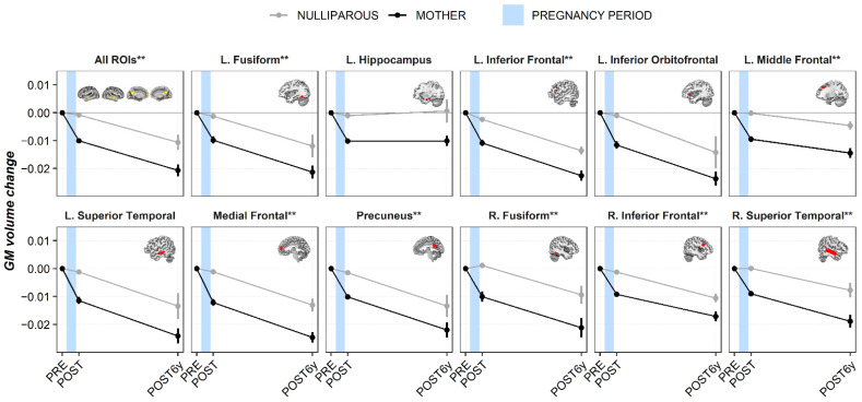
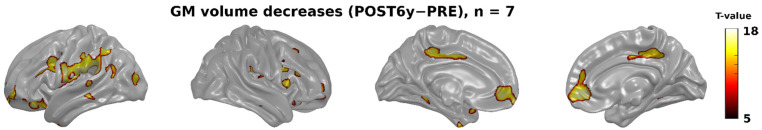
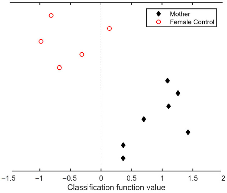
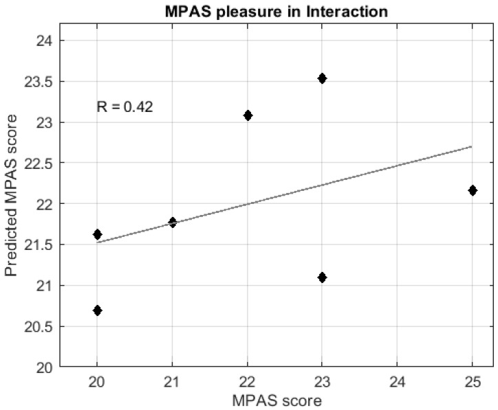
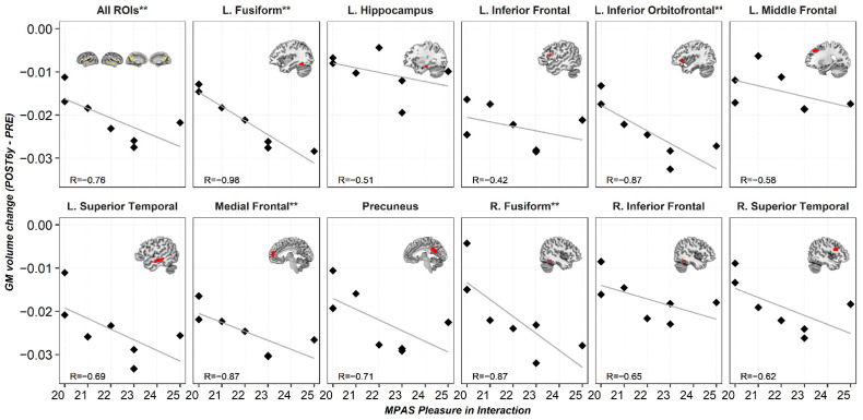
Neuroimaging researchers commonly assume that the brain of a mother is comparable to that of a nulliparous woman. However, pregnancy leads to pronounced gray matter volume reductions in the mother's brain, which have been associated with maternal attachment towards the baby. Beyond two years postpartum, no study has explored whether these brain changes are maintained or instead return to pre-pregnancy levels. The present study tested whether gray matter volume reductions detected in primiparous women are still present six years after parturition. Using data from a unique, prospective neuroimaging study, we compared the gray matter volume of 25 primiparous and 22 nulliparous women across three sessions: before conception (n = 25/22), during the first months of postpartum (n = 25/21), and at six years after parturition (n = 7/5). We found that most of the pregnancy-induced gray matter volume reductions persist six years after parturition (classifying women as having been pregnant or not with 91.67% of total accuracy). We also found that brain changes at six years postpartum are associated with measures of mother-to-infant attachment. These findings open the possibility that pregnancy-induced brain changes are permanent and encourage neuroimaging studies to routinely include pregnancy-related information as a relevant demographic variable.
Keywords: magnetic resonance imaging; maternal brain; neuroplasticity; postpartum; pregnancy.
Conflict of interest statement
The authors declare no conflict of interest. The funders had no role in the design of the study; in the collection, analyses, or interpretation of data; in the writing of the manuscript; or in the decision to publish the results.
Figures
Similar articles
-
The expectant brain-pregnancy leads to changes in brain morphology in the early postpartum period.Cereb Cortex. 2022 Sep 4;32(18):4025-4038. doi: 10.1093/cercor/bhab463. Cereb Cortex. 2022. PMID: 34942007 Free PMC article.
-
Time-sensitive changes in the maternal brain and their influence on mother-child attachment.Transl Psychiatry. 2024 Feb 9;14(1):84. doi: 10.1038/s41398-024-02805-2. Transl Psychiatry. 2024. PMID: 38331939 Free PMC article.
-
Becoming a mother entails anatomical changes in the ventral striatum of the human brain that facilitate its responsiveness to offspring cues.Psychoneuroendocrinology. 2020 Feb;112:104507. doi: 10.1016/j.psyneuen.2019.104507. Epub 2019 Nov 5. Psychoneuroendocrinology. 2020. PMID: 31757430
-
Pregnancy leads to long-lasting changes in human brain structure.Nat Neurosci. 2017 Feb;20(2):287-296. doi: 10.1038/nn.4458. Epub 2016 Dec 19. Nat Neurosci. 2017. PMID: 27991897
-
Physiology and Endocrinology Symposium: maternal immunological adjustments to pregnancy and parturition in ruminants and possible implications for postpartum uterine health: is there a prepartum-postpartum nexus?J Anim Sci. 2013 Apr;91(4):1639-49. doi: 10.2527/jas.2012-5934. Epub 2013 Jan 10. J Anim Sci. 2013. PMID: 23307838 Review.
Cited by
-
Enduring maternal brain changes and their role in mediating motherhood's impact on well-being.Sci Rep. 2024 Jul 18;14(1):16608. doi: 10.1038/s41598-024-67316-y. Sci Rep. 2024. PMID: 39025996 Free PMC article.
-
Sex differences in age-associated neurological diseases-A roadmap for reliable and high-yield research.Sci Adv. 2025 Mar 7;11(10):eadt9243. doi: 10.1126/sciadv.adt9243. Epub 2025 Mar 5. Sci Adv. 2025. PMID: 40043111 Free PMC article. Review.
-
Did Down-Regulated Instincts Enable Human Gene-Culture Coevolution?Evol Anthropol. 2025 Sep;34(3):e70015. doi: 10.1002/evan.70015. Evol Anthropol. 2025. PMID: 40811454 Free PMC article. Review.
-
Previous pregnancies might mitigate cortical brain differences associated with surgical menopause.Hum Brain Mapp. 2024 Jan;45(1):e26538. doi: 10.1002/hbm.26538. Epub 2023 Dec 8. Hum Brain Mapp. 2024. PMID: 38063284 Free PMC article.
-
The density of doublecortin cells in the piriform cortex is affected by transition to adulthood but not first pregnancy in mice.Brain Struct Funct. 2025 Jun 10;230(6):94. doi: 10.1007/s00429-025-02957-x. Brain Struct Funct. 2025. PMID: 40493077 Free PMC article.
References
-
- Lambert K.G., Kinsley C.H. Brain and behavioral modifications that accompany the onset of motherhood. Parenting. 2012;12:74–88. doi: 10.1080/15295192.2012.638868. - DOI
-
- Brunton P., Russell J. Maternal brain adaptations in pregnancy. In: Plant T., Zeleznik A., editors. Knobil and Neill’s Physiology of Reproduction: Two-Volume Set. Volume 2. Elsevier; Amsterdam, The Netherlands: 2015.
Grants and funding
- RTI2018-093952-B-100/Ministerio de Ciencia, Innovación y Universidades
- CP16/00096 PI17/00064/Instituto de Salud Carlos III
- ASPIDE 801091/European Union's Horizon 2020 research and innovation programme
- Miguel Servet Type I research contract CP16/00096 (Susanna Carmona)/Ministerio de Ciencia, Innovación y Universidades, Instituto de Salud Carlos III
- PFIS contract FI18/00255 (Magdalena Martínez-García)/Ministerio de Ciencia, Innovación y Universidades, Instituto de Salud Carlos III
- PEJD-2018-PRE/BMD-9401 (Maria Paternina-Díe)/Consejería de Educación e Investigación, Comunidad de Madrid, co-funded by European Social Fund "Investing in your future"
- LCF/PR/HR19/52160001/"la Caixa" Foundation
- SEV-2015-0505/CNIC is supported by the Ministerio de Ciencia, Innovación y Universidades and the Pro CNIC Foundation, and is a Severo Ochoa Center of Excellence
LinkOut - more resources
Full Text Sources
Other Literature Sources
