. 2021 Jan 28;22(3):1319.
doi: 10.3390/ijms22031319.

NLRP3 Inflammasome: A New Pharmacological Target for Reducing Testicular Damage Associated with Varicocele

Affiliations

NLRP3 Inflammasome: A New Pharmacological Target for Reducing Testicular Damage Associated with Varicocele

Pietro Antonuccio et al. Int J Mol Sci. .

Abstract

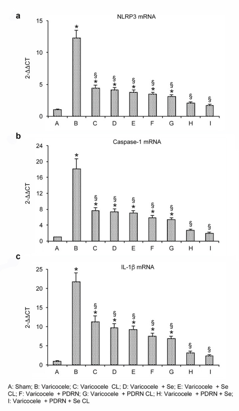
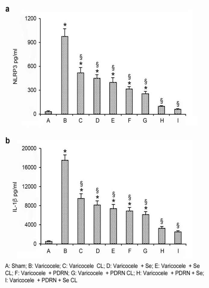
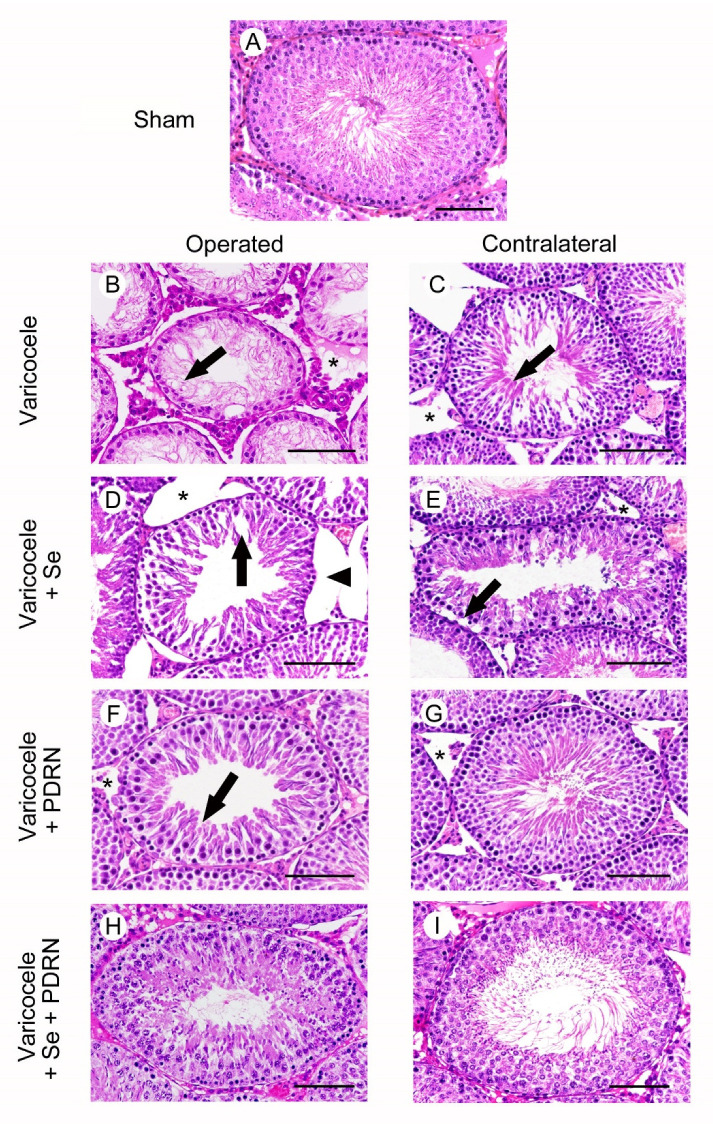
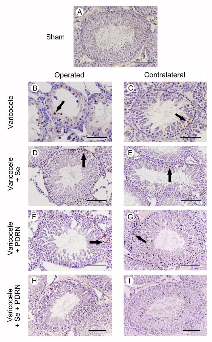
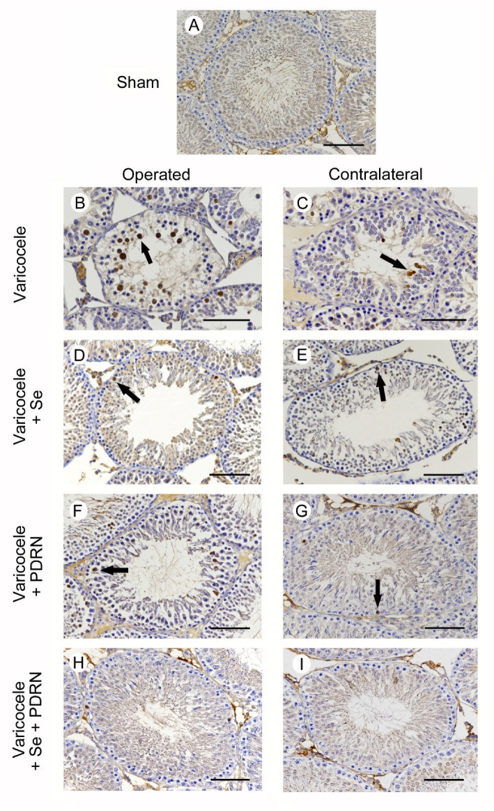
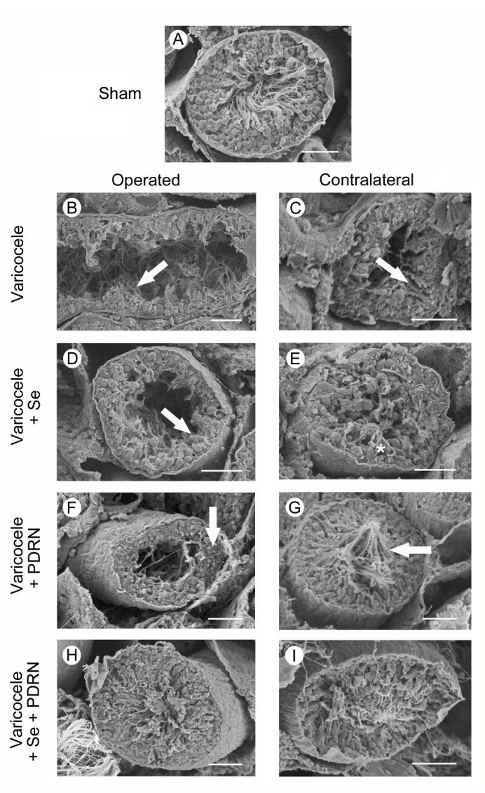
Many bioactive natural compounds are being increasingly used for therapeutics and nutraceutical applications to counteract male infertility, particularly varicocele. The roles of selenium and Polydeoxyribonucleotide (PDRN) were investigated in an experimental model of varicocele, with particular regard to the role of NLRP3 inflammasome. Male rats underwent sham operation and were daily administered with vehicle, seleno-L-methionine (Se), PDRN, and with the association Se-PDRN. Another group of rats were operated for varicocele. After twenty-eight days, sham and varicocele rats were sacrificed and both testes were weighted and analyzed. All the other rats were challenged for one month with the same compounds. In varicocele animals, lower testosterone levels, testes weight, NLRP3 inflammasome, IL-1β and caspase-1 increased gene expression were demonstrated. TUNEL assay showed an increased number of apoptotic cells. Structural and ultrastructural damage to testes was also shown. PDRN alone significantly improved all considered parameters more than Se. The Se-PDRN association significantly improved all morphological parameters, significantly increased testosterone levels, and reduced NLRP3 inflammasome, caspase-1 and IL-1β expression and TUNEL-positive cell numbers. Our results suggest that NLRP3 inflammasome can be considered an interesting target in varicocele and that Se-PDRN may be a new medical approach in support to surgery.

Keywords: A2A receptor; NLRP3 inflammasome; PDRN; nutraceuticals; selenium; testis; varicocele.


Conflict of interest statement

The authors declare no conflict of interest.

Figures

Figure 1
Figure 1
Real-time PCR analysis for NLRP3 (a), caspase-1 (b) and IL-1β (c) in the testes from rats of sham, varicocele, varicocele contralateral, varicocele plus Se (0.4 mg/kg/day i.p.), varicocele plus Se contralateral, varicocele plus PDRN (8 mg/kg/day i.p.), varicocele contralateral plus PDRN, varicocele plus Se plus PDRN, varicocele contralateral plus Se plus PDRN groups. * = p < 0.05 versus sham; § = p < 0.05 versus varicocele. Bars represent the mean ± SE of 7 experiments.
Figure 2
Figure 2
Quantitative evaluation of NLRP3 (a) and IL-1β (b) levels in the testes from rats of sham, varicocele, varicocele contralateral, varicocele plus Se (0.4 mg/kg/day i.p.), varicocele plus Se contralateral, varicocele plus PDRN (8 mg/kg/day i.p.), varicocele contralateral plus PDRN, varicocele plus Se plus PDRN, varicocele contralateral plus Se plus PDRN groups. * = p < 0.05 versus sham; § = p < 0.05 versus varicocele. Bars represent the mean ± SE of 7 experiments.
Figure 3
Figure 3
Structural organization of testes from rats of sham, varicocele, varicocele contralateral, varicocele plus Se (0.4 mg/kg/day i.p.), varicocele plus Se contralateral, varicocele plus PDRN (8 mg/kg/day i.p.), varicocele contralateral plus PDRN, varicocele plus Se plus PDRN, varicocele contralateral plus Se plus PDRN groups. (A): In sham testes, the seminiferous tubules have normal morphology. (B): Varicocele rats. The seminiferous epithelium shows reduced thickness with spermatogonia arranged on 1–2 rows and residual condensed sperm tails (arrow). In the extratubular compartment, a marked edema is evident (*). (C): Contralateral testes of the same group. The seminiferous tubules show many elongated spermatids (arrow). The extratubular compartment is edematous and hyperemic (*). (D): In varicocele operated rats treated with Se, the seminiferous epithelium is detached from the basement membrane (arrowhead) and many large clefts are present in its wall (arrow). The extratubular compartment is enlarged (*). (E): In the contralateral testes of the same group, peripheral spermatogonia are often detached from the basement membrane (arrow). The extratubular compartment shows hyperemic blood vessels and edema (*). (F): Varicocele operated rats treated with PDRN. The seminiferous epithelium shows intercellular clefts among round or elongated spermatids (arrow). A mild edema is present in the extratubular compartment (*). (G): Contralateral testes of the same group. The seminiferous epithelium has many elongated spermatids and mature spermatozoa; the extratubular compartment shows only a mild edema (*). (H,I): In the seminiferous tubules of both operated and contralateral testes of varicocele rats treated with Se plus PDRN, the seminiferous epithelium is close to normal. (Scale bar = 50 µm).
Figure 4
Figure 4
Assessment of apoptosis in the testes with TUNEL staining technique from rats of sham, varicocele, varicocele contralateral, varicocele plus Se (0.4 mg/kg/day i.p.), varicocele plus Se contralateral, varicocele plus PDRN (8 mg/kg/day i.p.), varicocele contralateral plus PDRN, varicocele plus Se plus PDRN, varicocele contralateral plus Se plus PDRN groups. (A): In sham rats, no TUNEL positive cells can be observed. (B): In the testes of varicocele rats, a large number of TUNEL-positive germ cells (arrow), placed along the wall of the tubules, are observed. (C): In the contralateral testes of the same rats, some isolated TUNEL-positive germ cells are present in the peripheral part of the seminiferous tubules (arrow). (D): In varicocele rats treated with Se, many TUNEL-positive cells are present in the periphery of the seminiferous tubules (arrow). (E): In the contralateral testes of the same group, some TUNEL-positive cells are located in the external part of the tubules (arrow). (F): In the testes of varicocele rats treated with PDRN, the number of TUNEL-positive cells is reduced, and they are located along the peripheral part of the tubules (arrow). (G): In contralateral testes of the same group, only a few TUNEL-positive cells (arrow) are present in the seminiferous epithelium. H, I: In the seminiferous tubules of both operated and contralateral testes of varicocele rats treated with Se plus PDRN, very rare or no TUNEL-positive cells are observed. (Scale bar: 50 µm).
Figure 5
Figure 5
Immunohistochemical localization of caspase-1 in the testes from rats of sham, varicocele, varicocele contralateral, varicocele plus Se (0.4 mg/kg/day i.p.), varicocele plus Se contralateral, varicocele plus PDRN (8 mg/kg/day i.p.), varicocele contralateral plus PDRN, varicocele plus Se plus PDRN, varicocele contralateral plus Se plus PDRN groups. (A): In sham rats, no caspase-1 positive cells can be observed. (B): In varicocele rats, a large number of caspase-1 positive cells are located in the wall of the tubules (arrow). (C): In the contralateral testes of the same rats, some isolated caspase-1 positive cells are present (arrow). (D): In varicocele rats treated with Se, many caspase-1 positive cells are present in the periphery of the seminiferous tubules (arrow). (E): In the contralateral testes of the same group, some caspase-1 positive cells are located in the external part of the tubules (arrow). (F): In the testes of varicocele rats treated with PDRN, fewer caspase-1 positive cells are located along the periphery of the tubules (arrow). (G): In contralateral testes of the same group, only isolated caspase-1 positive cells (arrow) are present. (H,I): In the seminiferous tubules of both operated and contralateral testes of varicocele rats treated with Se plus PDRN, very rare or no caspase-1 positive cells are demonstrated. (Scale bar: 50 µm).
Figure 6
Figure 6
Scanning electron micrographs of testes from sham, varicocele, varicocele contralateral, varicocele plus Se (0.4 mg/kg/day i.p.), varicocele plus Se contralateral, varicocele plus PDRN (8 mg/kg/day i.p.), varicocele contralateral plus PDRN, varicocele plus Se plus PDRN, varicocele contralateral plus Se plus PDRN groups. (A): Sham animals. Note the normal structure of the seminiferous tubules. (B): Varicocele rats. Tubules show evident reduction in their height and condensed sperm tails (arrow). (C): In the contralateral testes of the same group, some clefts are present in the seminiferous epithelium (arrow). (D): In varicocele rats treated with Se, a low and irregularly arranged seminiferous epithelium is evident (arrow). (E): In the contralateral testes of the same group, only a few spermatozoa can be observed (asterisk). (F): In varicocele rats treated with PDRN the tubular lumen is reduced, owing to the presence of a higher epithelium (arrow). (G): In the contralateral testes of the same group, many spermatozoa are present (arrow). (H,I): In varicocele rats treated with Se plus PDRN, both the operated and the contralateral testes show close to normal organization. (Scale bar: 50 μm).

References

    1. Cho C.L., Esteves S.C., Agarwal A. Novel insights into the pathophysiology of varicocele and its association with reactive oxygen species and sperm DNA fragmentation. Asian J. Androl. 2016;18:186–193. doi: 10.4103/1008-682X.170441. - DOI - PMC - PubMed
    1. Alsaikhan B., Alrabeeah K., Delouya G., Zini A. Epidemiology of varicocele. Asian J. Androl. 2016;18:179–181. doi: 10.4103/1008-682X.172640. - DOI - PMC - PubMed
    1. Hassanin A.M., Ahmed H.H., Kaddah A.N. A global view of the pathophysiology of varicocele. Andrology. 2018;6:654–661. doi: 10.1111/andr.12511. - DOI - PubMed
    1. Pastuszak A.W., Wang R. Varicocele and testicular function. Asian J. Androl. 2015;17:659–667. doi: 10.4103/1008-682X.153539. - DOI - PMC - PubMed
    1. Barati E., Nikzad H., Karimian M. Oxidative stress and male infertility: Current knowledge of pathophysiology and role of antioxidant therapy in disease management. Cell. Mol. Life. Sci. 2020;77:93–113. doi: 10.1007/s00018-019-03253-8. - DOI - PMC - PubMed

MeSH terms

LinkOut - more resources