. 2021 Jul 21;32(7):1374-1392.
doi: 10.1021/acs.bioconjchem.0c00691. Epub 2021 Feb 1.

Structural Investigations, Cellular Imaging, and Radiolabeling of Neutral, Polycationic, and Polyanionic Functional Metalloporphyrin Conjugates

Affiliations

Structural Investigations, Cellular Imaging, and Radiolabeling of Neutral, Polycationic, and Polyanionic Functional Metalloporphyrin Conjugates

Valeria Ciaffaglione et al. Bioconjug Chem. .

Abstract

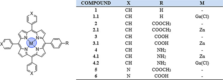
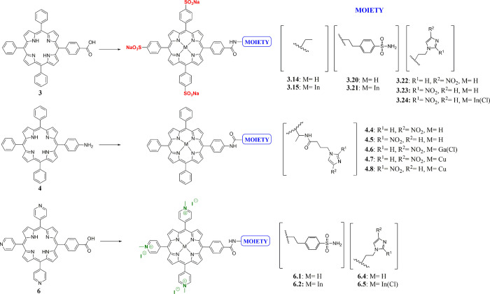
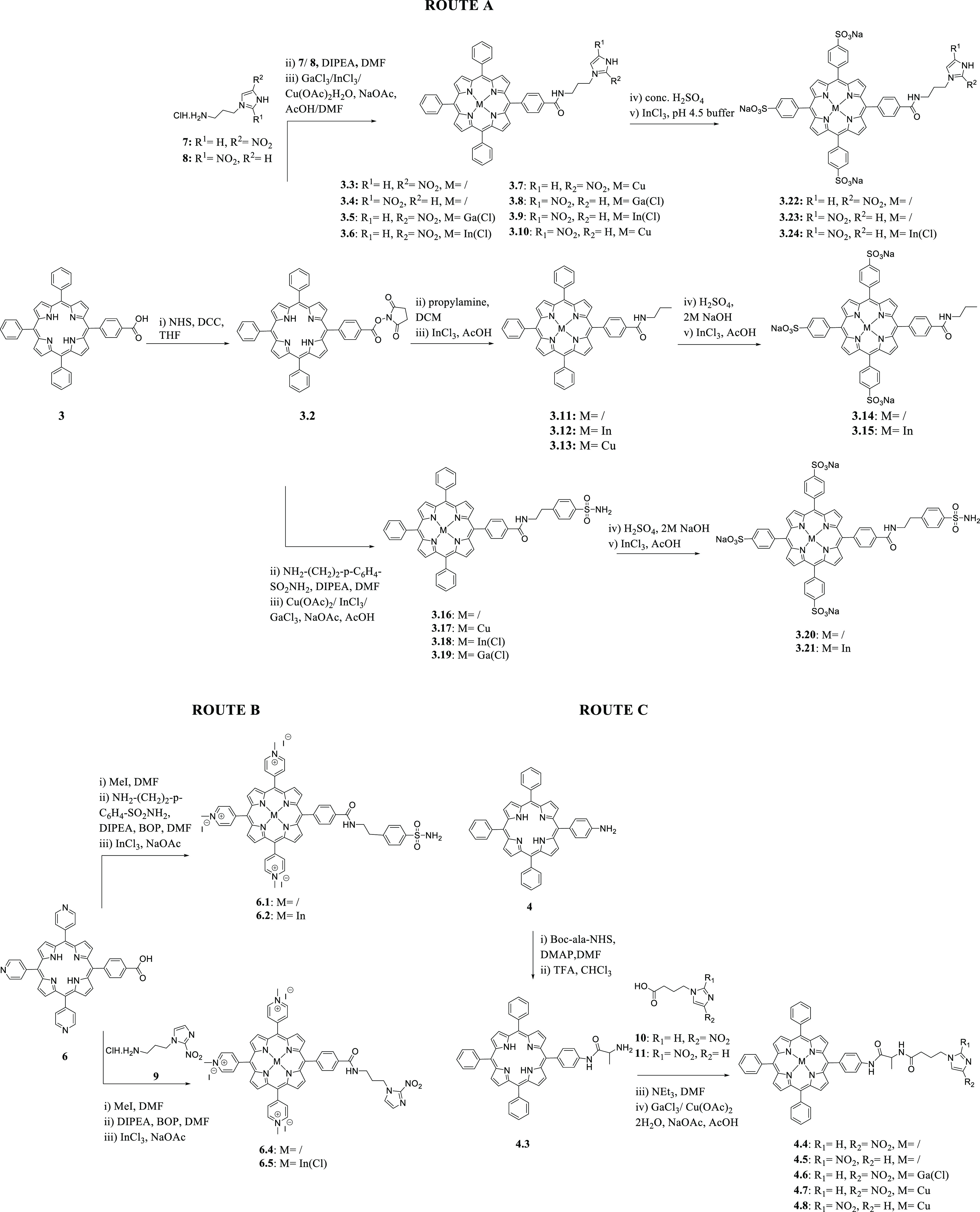
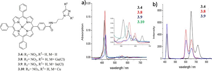
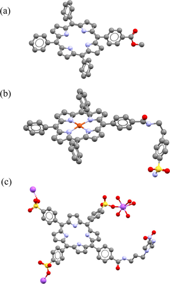
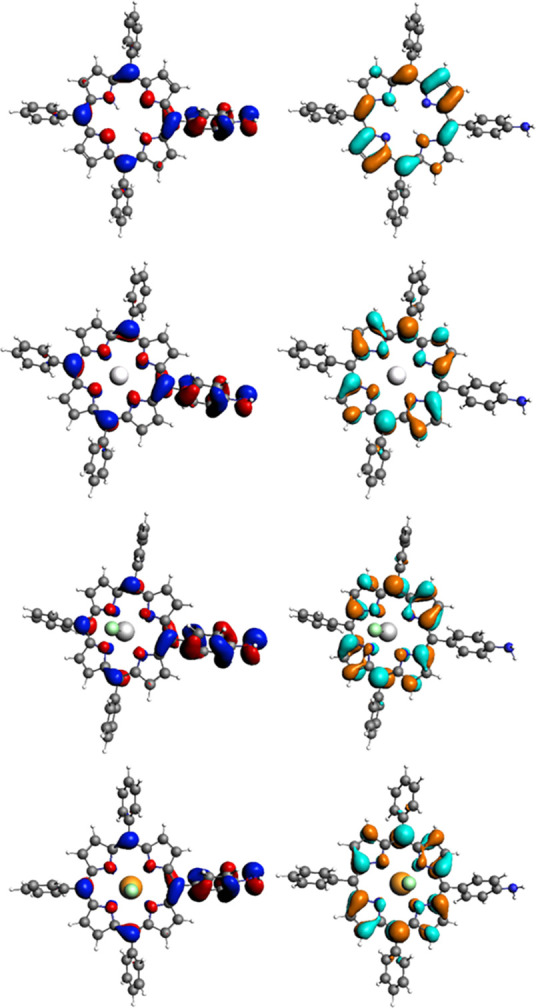
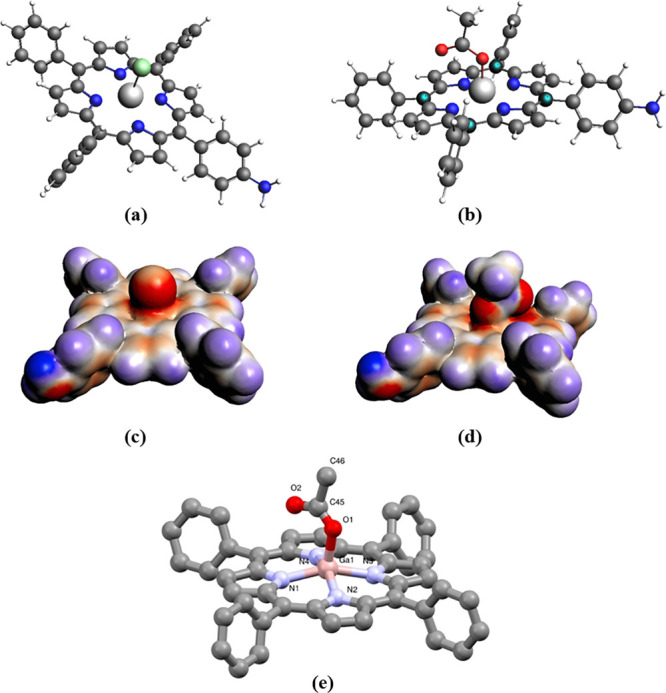
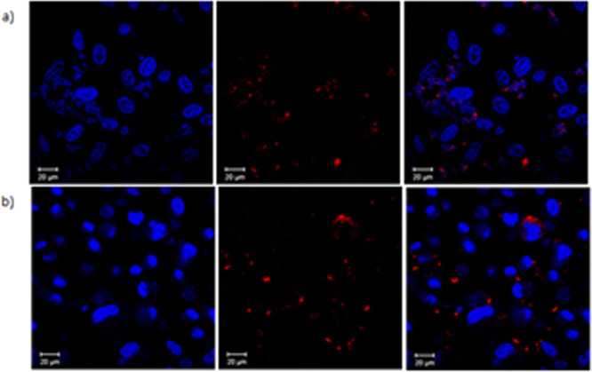
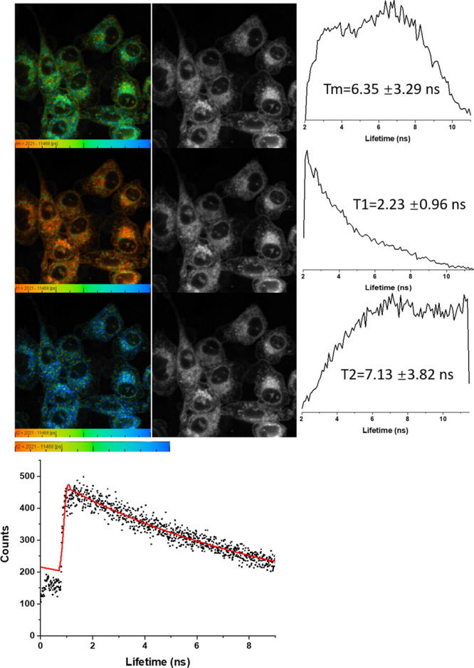
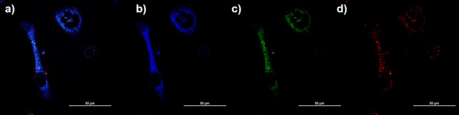
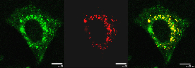
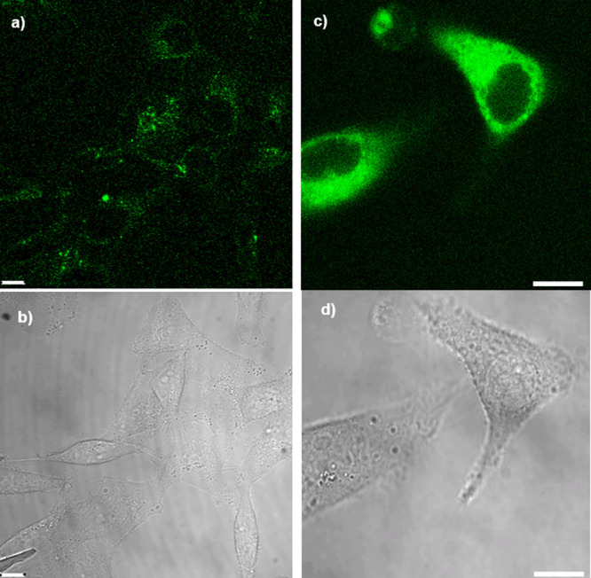
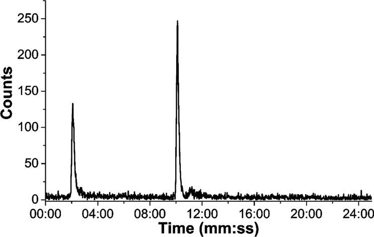
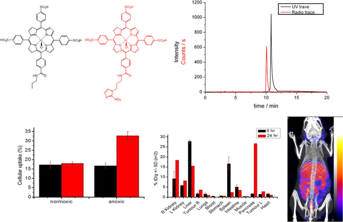
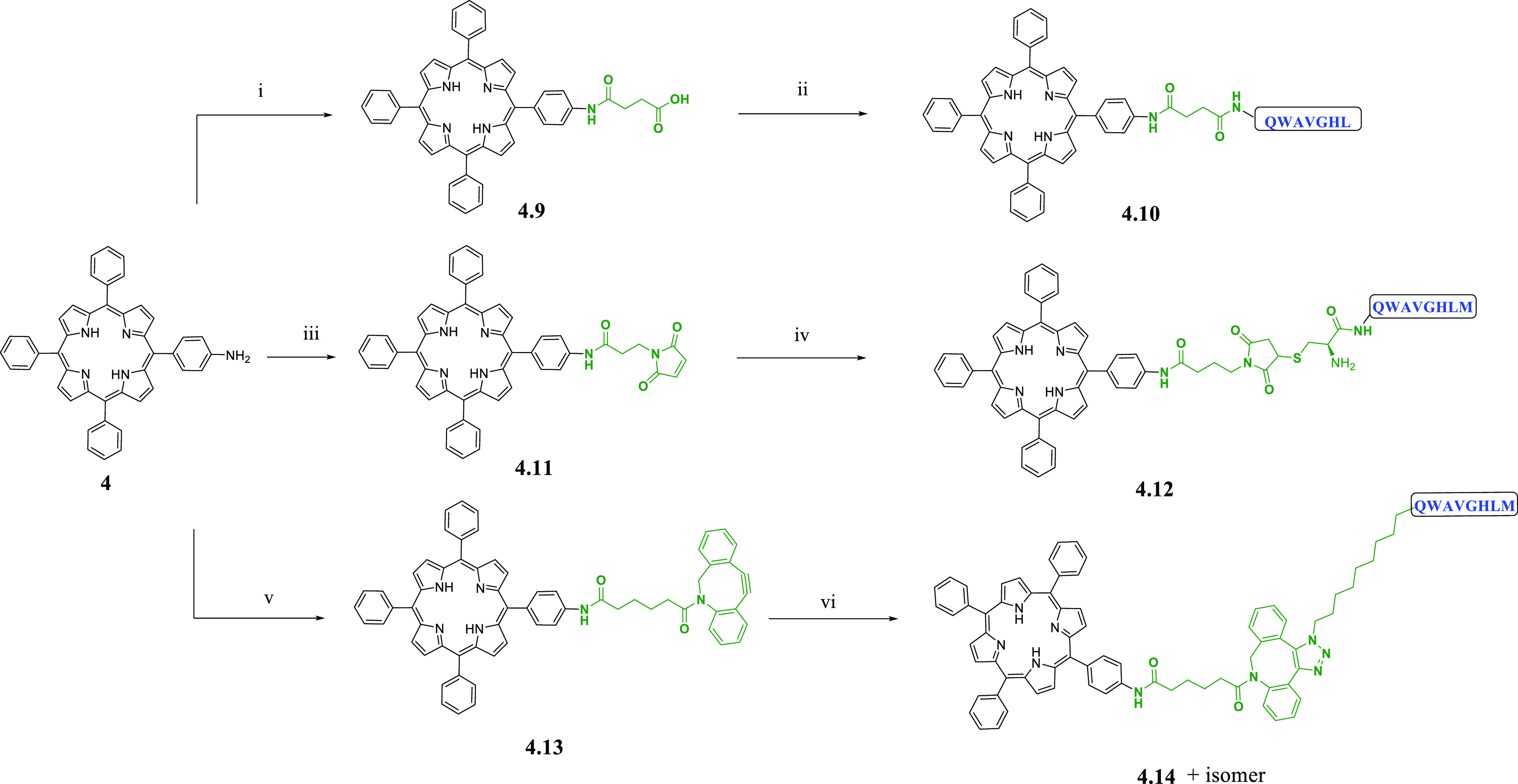
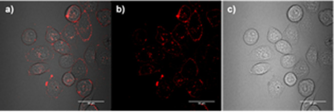
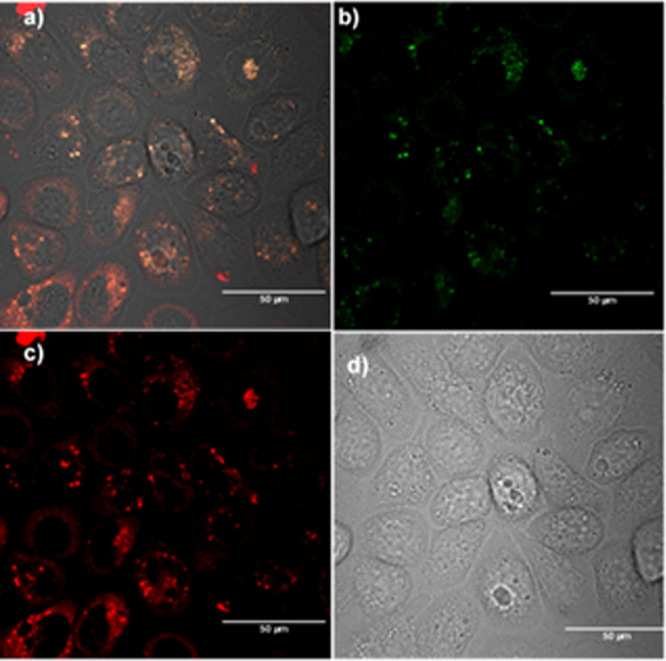
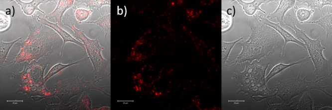
Over the past decade, porphyrin derivatives have emerged as invaluable synthetic building blocks and theranostic kits for the delivery of cellular fluorescence imaging and photodynamic therapy. Tetraphenylporphyrin (TPP), its metal complexes, and related derivatives have been investigated for their use as dyes in histology and as components of multimodal imaging probes. The photophysical properties of porphyrin-metal complexes featuring radiometals have been a focus of our attention for the realization of fluorescence imaging probes coupled with radioimaging capabilities and therapeutic potential having "true" theranostic promise. We report hereby on the synthesis, radiochemistry, structural investigations, and preliminary in vitro and in vivo uptake studies on a range of functionalized porphyrin-based derivatives. In pursuit of developing new porphyrin-based probes for multimodality imaging applications, we report new functionalized neutral, polycationic, and polyanionic porphyrins incorporating nitroimidazole and sulfonamide moieties, which were used as targeting groups to improve the notoriously poor pharmacokinetics of porphyrin tags. The resulting functional metalloporphyrin species were stable under serum challenges and the nitroimidazole and sulfonamide derivatives remained fluorescent, allowing in vitro confocal studies and visualization of the lysosomal uptake in a gallium(III) sulfonamide derivative. The molecular structures of selected porphyrin derivatives were determined by single crystal X-ray diffraction using synchrotron radiation. We also investigated the nature of the emission/excitation behavior of model functional porphyrins using in silico approaches such as TD DFT in simple solvation models. The conjugation of porphyrins with the [7-13] and [7-14] fragments of bombesin was also achieved, to provide targeting of the gastrin releasing peptide receptor (GRPR). Depending on the metal, probe conjugates of relevance for single photon emission computed tomography (SPECT) or positron emission tomography (PET) probes have been designed and tested hereby, using TPP and related functional free base porphyrins as the bifunctional chelator synthetic scaffold and 111In[In] or 68Ga[Ga], respectively, as the central metal ions. Interestingly, for simple porphyrin conjugates good radiochemical incorporation was obtained for both radiometals, but the presence of peptides significantly diminished the radio-incorporation yields. Although the gallium-68 radiochemistry of the bombesin conjugates did not show radiochemical incorporation suitable for in vivo studies, likely because the presence of the peptide changed the behavior of the TPP-NH2 synthon taken alone, the optical imaging assays indicated that the conjugated peptide tags do mediate uptake of the porphyrin units into cells.


Conflict of interest statement

The authors declare no competing financial interest.

Figures

Figure 1
Figure 1
Structural representation of the basic porphyrin-based building blocks: “cold” metal complexes and their precursors.
Figure 2
Figure 2
Chemical structures of the most representative synthesized polyanionic, neutral and polycationic porphyrins, and their precursors.
Scheme 1
Scheme 1. Synthetic Scheme of Functionalized Porphyrin Species
Full experimental details and characterization data are given in SI.
Figure 3
Figure 3
Structural representation of compounds 3.4 and 3.83.10. (a) Overlay of UV–vis spectra of 3.4 (black), 3.8 (red), 3.9 (blue), and 3.10 (green) (all spectra measured at 0.2 μM in DMF) with expansion of the Q region inset. (b) Overlay of fluorescence spectra of 3.4, 3.8, and 3.9 with λex at 418 nm (all spectra were measured at 0.2 μM in DMF).
Figure 4
Figure 4
Molecular structures determined from single crystal X-ray diffraction for compounds 2 (a), 3.17 (b), and 3.22 (c). Color code: magenta = Na, blue = N, yellow = S, red = O, gray = C. H atoms and disordered units have been removed. Crystallography details for all structures are given in the SI.
Figure 5
Figure 5
Illustrations of significant frontier orbitals: HOMO (left, blue/red) and LUMO (right, orange/cyan) of 4, 4.1, 4.2, and 4-InCl (top to bottom). Further images are given in SI.
Figure 6
Figure 6
Optimized geometries for model compounds: (a) 4.2-Cl substituted and (b) 4.2-OAc substituted; corresponding COSMO surfaces: (c) 4.2-Cl substituted and (d) 4.2-OAc models; the molecular structure determined from synchrotron single crystal X-ray diffraction analysis for compound 1.1-OAc substituted (e).
Figure 7
Figure 7
Confocal images of TPP-COOMe (2) and TPP-COOH (3) at 10 μM solution in DMSO (1%) with PC-3 cells after incubation for 1 h at 37 °C. Hoechst nuclear stain was used (1 μg/mL, blue) to visualize the cell nuclei and porphyrins show red fluorescence: (a) Hoechst stained nuclei, porphyrin 2, overlay of blue and red emission channels images; (b) Hoechst stained nuclei, porphyrin 3, overlay of blue and red emission images; Scale bar 20 μm.
Figure 8
Figure 8
2-Photon FLIM of compound 3 in PC-3 cells after 20 min incubation, 10 μM total conc., DMSO (1%), 3 mW, 810 nm excitation (top three rows). The corresponding solution TCSPC spectra and fitted curve are given: 10 μM in DMSO, χ2 1.65, and τ1 20 ps (38%, indicating aggregation, forming short-lived excimers with lifetime within instrument response) and a porphyrin-characteristic τ2 of 11.37 ns (62%).
Figure 9
Figure 9
Confocal images for compound 4 in PC-3 cells incubated for 12 h at 10 μm concentration, 37 °C: (a) overlay of blue-green-red channels; (b) blue channel (λem = 417–477 nm); (c) green channel (λem = 500–550 nm); (d) red channel (λem = 570–750 nm) (see SI for additional images and details).
Figure 10
Figure 10
In vitro confocal fluorescence uptake studies of 4.6 (10 μM) in HCT116 (CAIX positive) living cells (4 h incubation, 37 °C, λex = 405 nm, λem = 600 nm); (a) 4.6 uptake; (b) Lysotracker Red uptake; (c) overlay of images recorded after Lysotracker Red dye staining and 4.6 uptake (scale bar 10 μm).
Figure 11
Figure 11
(a) Confocal fluorescence imaging in living CA IX positive HCT116 cells of the free base precursor 3.16 10 μM 16 h (λex = 410 nm, λem = 625 nm), and (b) corresponding bright field image. (c) Confocal fluorescence imaging in living CA IX positive HCT116 cells of the gallium complex 3.19 16 h, λex = 405 nm, λem = 625 nm and (d) corresponding bright field image; Scale bar = 10 μm.
Figure 12
Figure 12
Radiochromatogram of compound [68Ga]4.2 labeled with generator-produced [68Ga]GaCl3 ions in water, after purification by chromatography over a C18-silica cartridge.
Figure 13
Figure 13
(top) Structures of [111In]3.24 (red) and [111In]3.15 (black) with corresponding UV–vis and radio HPLC traces. (bottom) Hypoxia selectivity assay in vitro (HeLa cell line), biodistribution data of [111In]3.15, and SPECT image measured at 24 h of MKN45 tumor bearing mice injected with 3.15 (n = 6). Further details are given in SI.
Scheme 2
Scheme 2. Coupling Reaction Scheme of Functionalized Porphyrin-Based Conjugates with Peptides
Reagents and conditions: (i) succinic anhydride, TEA, CHCl3; (ii) PyBOP, DIPEA, DMF, BBN[7-13]; (iii) 3-maleimidopropionic acid, HOBt, EDC·HCl, DCM; (iv) Cys-BBN[7-14]-NH2, pyridine, DMSO; (v) dibenzocyclooctyne acid, HOBt, EDC·HCl, DIPEA, DCM; (vi) 11-azido undecanoyl-BBN[7-14]-NH2, DMSO.
Figure 14
Figure 14
Confocal imaging of the butanoic acid linker-conjugated compound 4.9 (50 μM, 1% DMSO) in PC3 cells after incubation (20 min, 37 °C): λex = 488 nm, λ = 650–700 nm; Scale bar 50 μm.
Figure 15
Figure 15
Confocal imaging of the peptide-conjugated compound 4.10 (200 μM, 1% DMSO) in PC3 cells after incubation (20 min, 37 °C). λex = 488 nm, λ = 650–700 nm. Scale bar 50 μm.
Figure 16
Figure 16
Confocal imaging of the peptide-conjugated compound 4.12 (10 μM, 1% DMSO) in PC3 cells after incubation (20 min, 37 °C). λex = 488 nm, λ = 650–700 nm. Scale bar 20 μm.

References

    1. Dolphin D.The Porphyrins Volume VII: Biochemistry, part B.
    1. Urata G. (1995) [The chemistry of porphyrins and their precursors on the heme biosynthetic chain]. Nihon rinsho 53 (6), 1319–28. - PubMed
    1. Josefsen L. B.; Boyle R. W. (2012) Unique diagnostic and therapeutic roles of porphyrins and phthalocyanines in photodynamic therapy, imaging and theranostics. Theranostics 2 (9), 916–66. 10.7150/thno.4571. - DOI - PMC - PubMed
    1. Higashino T.; Imahori H. (2015) Porphyrins as excellent dyes for dye-sensitized solar cells: recent developments and insights. Dalton transactions 44 (2), 448–63. 10.1039/C4DT02756F. - DOI - PubMed
    1. Yan G. P.; Bischa D.; Bottle S. E. (2007) Synthesis and properties of novel porphyrin spin probes containing isoindoline nitroxides. Free Radical Biol. Med. 43 (1), 111–6. 10.1016/j.freeradbiomed.2007.03.032. - DOI - PubMed

Publication types

LinkOut - more resources