Review
. 2021 Dec;13(2_suppl):367S-374S.
doi: 10.1177/1947603520988770. Epub 2021 Feb 1.

A Review of the Collagen Orientation in the Articular Cartilage

Affiliations
Review

A Review of the Collagen Orientation in the Articular Cartilage

Roy D Bloebaum et al. Cartilage. 2021 Dec.

Abstract

Objective: There has been a debate as to the alignment of the collagen fibers. Using a hand lens, Sir William Hunter demonstrated that the collagen fibers ran perpendicular and later aspects were supported by Benninghoff. Despite these 2 historical studies, modern technology has conflicting data on the collagen alignment.

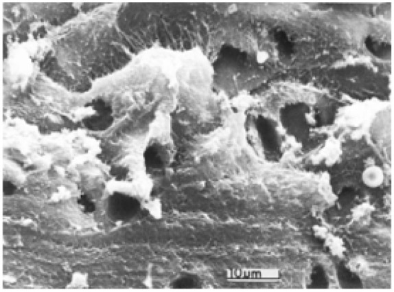
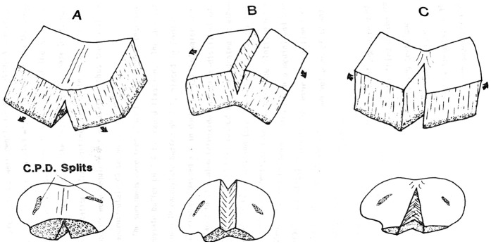
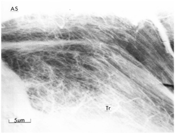
Design: Ten mature New Zealand rabbits were used to obtain 40 condyle specimens. The specimens were passed through ascending grades of alcohol, subjected to critical point drying (CPD), and viewed in the scanning electron microscope. Specimens revealed splits from the dehydration process. When observing the fibers exposed within the opening of the splits, parallel fibers were observed to run in a radial direction, normal to the surface of the articular cartilage, radiating from the deep zone and arcading as they approach the surface layer. After these observations, the same samples were mechanically fractured and damaged by scalpel.

Results: The splits in the articular surface created deep fissures, exposing parallel bundles of collagen fibers, radiating from the deep zone and arcading as they approach the surface layer. On higher magnification, individual fibers were observed to run parallel to one another, traversing radially toward the surface of the articular cartilage and arcading. Mechanical fracturing and scalpel damage induced on the same specimens with the splits showed randomly oriented fibers.

Conclusion: Collagen fiber orientation corroborates aspects of Hunter's findings and compliments Benninghoff. Investigators must be aware of the limits of their processing and imaging techniques in order to interpret collagen fiber orientation in cartilage.

Keywords: articular cartilage; collagen alignment; review; scanning electron microscopy; tissue.


Conflict of interest statement

Declaration of Conflicting Interests: The author(s) declared no potential conflicts of interest with respect to the research, authorship, and/or publication of this article.

Figures

Figure 1.
Figure 1.
A high-powered view of the random fibers, as seen in the deep, middle/transversal, and superficial zone of the articular cartilage, created as a result of the mechanical fracture technique.
Figure 2.
Figure 2.
A high-powered view of the disrupted fibers traversing the 3 zones from scalpel damage. Empty lacunae can be seen.
Figure 3.
Figure 3.
The steps taken to collect specimens with spontaneously occurring splits observed following tissue processing, due to shrinkage.
Figure 4.
Figure 4.
Numerous bundles with parallel fibers traversing from the deep and middle zones of the cartilage to the surface zone. This figure demonstrates the irregularly shaped splits that formed as a result of shrinkage. One could imagine that if the split region were reassembled there would be no gaps between the bundles.
Figure 5.
Figure 5.
A diagram of the fracture technique, following Clarke’s 1971 protocol. The specimens were prepared to retain the calcified zone and remnants of the subchondral bone to assure that the 3 zones (deep, middle, and superficial) were preserved.
Figure 6.
Figure 6.
A low-powered view of the scalpel damage. This damage was incomplete in this specimen, allowing the remainder of the tissue to split open, exposing the deep zone parallel fiber bundles, undamaged by the blade. The articular surface (AS), the scalpel region of damage (EX) shows the pattern of the edge of the scalpel blade, and the split edge (CP).
Figure 7.
Figure 7.
A high-powered view of the continuous parallel fibers communicating to the surface.
Figure 8.
Figure 8.
Arcading parallel fibers (Tr) bending toward the articular surface (AS).
Figure 9.
Figure 9.
A mechanically fractured surface, revealing random fibers and bundles along the depth of the articular cartilage structure from deep zone, middle zone, and superficial zone to the surface. Chondrocyte lacunae (CL) can be observed.
Figure 10.
Figure 10.
Arcading fiber bundles observed in the mechanically fractured specimen, near the surface layer.
Figure 11.
Figure 11.
A low-powered view of scalpel damage showing striation pattern from the scalpel blade edge (EX) traversing the 3 zones. The articular surface is represented by AS.

Similar articles

Cited by

References

    1. Hunter WH. Of the structure and diseases of articular cartilage. Philos Trans R Soc Lond B Biol Sci. 1742;42:514-521.
    1. Benninghoff A. Form und bau der Gelenknorpel in ihren Bezeihungen zur Funktion [in German]. Z Zellforsch Mikrosk Anat. 1925;2:783-825.
    1. Clarke IC. Human articular surface contours and related surface depression frequency studies. Ann Rheum Dis. 1971;30:15-23. doi:10.1136/ard.30.1.15 - DOI - PMC - PubMed
    1. Jeffery AK, Blunn GW, Archer CW, Bentley G. Three-dimensional collagen architecture in bovine articular cartilage. J Bone Joint Surg Br. 1991;73:795-801. - PubMed
    1. Hunziker EB, Michel M, Studer D. Ultrastructure of adult human articular cartilage matrix after cryotechnical processing. Microsc Res Tech. 1997;37:271-284. doi:10.1002/(sici)1097-0029(19970515)37:43.0.co;2-of - DOI - PubMed

Publication types