. 2021 Feb 1;18(1):6.
doi: 10.1186/s12989-021-00398-y.

Macrophage autophagy protects mice from cerium oxide nanoparticle-induced lung fibrosis

Affiliations

Macrophage autophagy protects mice from cerium oxide nanoparticle-induced lung fibrosis

Balasubramanyam Annangi et al. Part Fibre Toxicol. .

Abstract

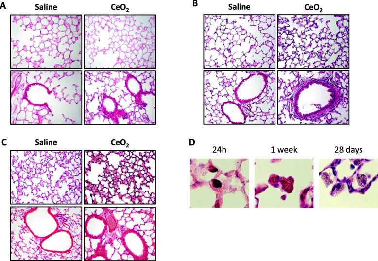
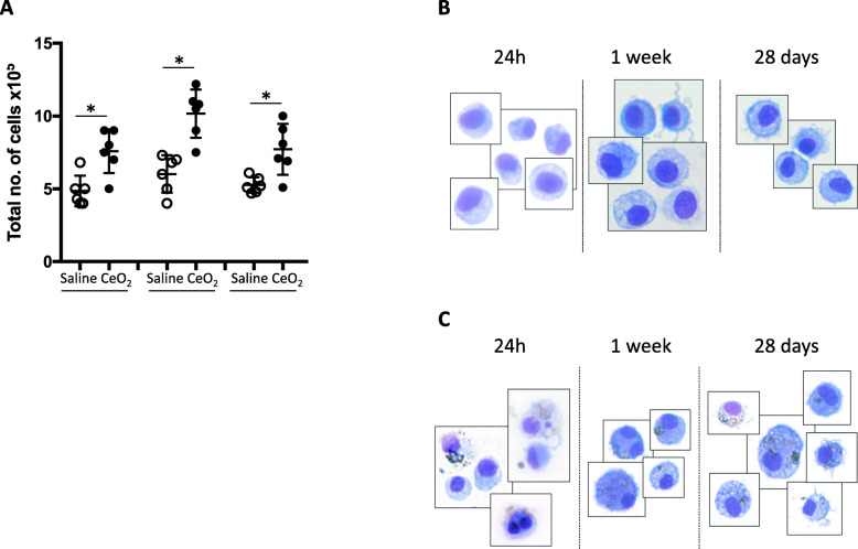
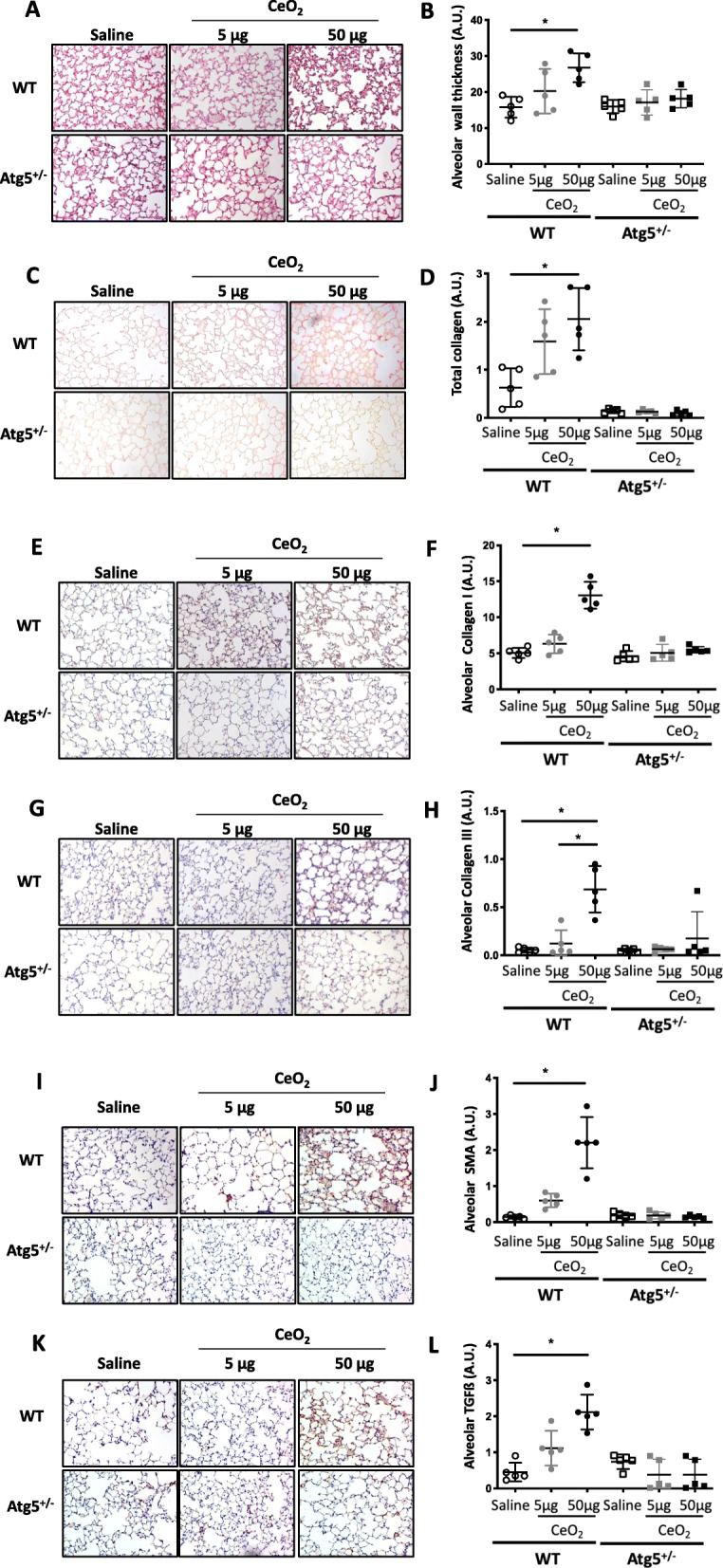
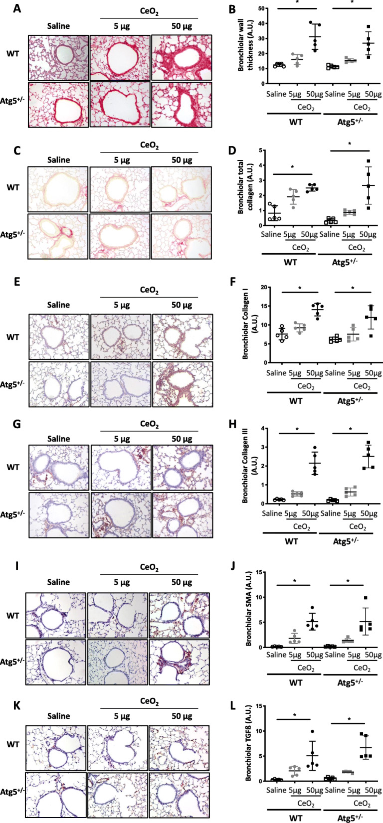
Background: Cerium (Ce) is a rare earth element, rapidly oxidizing to form CeO2, and currently used in numerous commercial applications, especially as nanoparticles (NP). The potential health effects of Ce remain uncertain, but literature indicates the development of rare earth pneumoconiosis accompanied with granuloma formation, interstitial fibrosis and inflammation. The exact underlying mechanisms are not yet completely understood, and we propose that autophagy could be an interesting target to study, particularly in macrophages. Therefore, the objective of our study was to investigate the role of macrophagic autophagy after pulmonary exposure to CeO2 NP in mice. Mice lacking the early autophagy gene Atg5 in their myeloid lineage and their wildtype counterparts were exposed to CeO2 NP by single oropharyngeal administration and sacrificed up to 1 month after. At that time, lung remodeling was thoroughly characterized (inflammatory cells infiltration, expression of fibrotic markers such as αSMA, TGFβ1, total and type I and III collagen deposition), as well as macrophage infiltration (quantification and M1/M2 phenotype).

Results: Such pulmonary exposure to CeO2 NP induces a progressive and dose-dependent lung fibrosis in the bronchiolar and alveolar walls, together with the activation of autophagy. Blockage of macrophagic autophagy protects from alveolar but not bronchiolar fibrosis, via the modulation of macrophage polarization towards M2 phenotype.

Conclusion: In conclusion, our findings bring novel insight on the role of macrophagic autophagy in lung fibrogenesis, and add to the current awareness of pulmonary macrophages as important players in the disease.

Keywords: Alveolar fibrosis - autophagy - macrophage polarization; Nanoparticle.


Conflict of interest statement

No competing interest to declare.

Figures

Fig. 1
Fig. 1
Exposure to CeO2 NP induces a progressive lung remodeling. Representative lung tissue sections of C57Bl/6 mice exposed to Saline or CeO2 NP and stained with Hematoxylin Eosin (HE) after 1 day (Panel a), 7 days (Panel b) or 28 days (Panel c). Original magnification × 200. Panel d: higher magnification of alveolar macrophages with internalized NP
Fig. 2
Fig. 2
Exposure to CeO2 NP induces an increase in BAL cellularity. Quantification of total cell count in BAL fluid (Panel a). Each individual circle represents the value obtained from one animal (empty circle: saline exposure – plain circle: CeO2 NP-exposure). *p < 0.05. Typical images of BAL macrophages from Saline-exposed (Panel b) or CeO2 NP-exposed (Panel c) mice sacrifice after 24 h, 1 week or 28 days
Fig. 3
Fig. 3
Exposure to CeO2 NP induces autophagy. Representative lung tissue sections of GFP-LC3 mice exposed to Saline or CeO2 NP after 1 day (Panel a), 7 days (Panel b) or 28 days (Panel c). Original magnification × 200. Panel d: representative lung tissue sections of C57Bl/6 mice exposed to Saline or 50 μg CeO2 NP and stained, after 28 days, for Atg5. Original magnification × 200
Fig. 4
Fig. 4
Atg5+/− mice are protected from alveolar remodeling. Representative lung tissue sections of WT and Atg5+/− mice exposed to Saline or CeO2 NP (5 or 50 μg, observation at 28 days), after HES (Panel a and b for quantification), Sirius red (Panel c and d for quantification), Collagen type I (Panel e and f for quantification), Collagen type III (Panel g and h for quantification), αSMA (Panel i and j for quantification) and TGF-ß (Panel k and l for quantification). Original magnification × 200. Each individual circle represents the mean value obtained from one animal. Empty circle: saline exposure. Grey circle: 5 μg CeO2 NP exposure. Plain circle: 50 μg CeO2 NP exposure. *p < 0.05
Fig. 5
Fig. 5
Atg5+/− mice are not protected from bronchiolar remodeling. Representative lung tissue sections of WT and Atg5+/− mice exposed to Saline or CeO2 NP (5 or 50 μg, observation at 28 days), after HES (Panel a and b for quantification), Sirius red (Panel c and d for quantification), Collagen type I (Panel e and f for quantification), Collagen type III (Panel g and h for quantification), αSMA (Panel i and j for quantification) and TGF-ß (Panel k and l for quantification). Original magnification × 200. Each individual circle represents the mean value obtained from one animal. Empty circle: saline exposure. Grey circle: 5 μg CeO2 NP exposure. Plain circle: 50 μg CeO2 NP exposure. *p < 0.05
Fig. 6
Fig. 6
Characterization of CeO2-induced macrophage recruitment and polarization in mice. Representative lung tissue sections of C57Bl/6 mice exposed to Saline or CeO2 NP (5 or 50 μg, observation at 28 days), after immunostaining for CD107b, as a marker of total number of macrophages (Panel a). Original magnification × 200. Quantification of CD107b positive cells (Panel b). Representative lung tissue sections of mice exposed to Saline or CeO2 NP (5 or 50 μg, observation at 28 days), after immunostaining for CD80 (Panel c and d for quantification), CD68 (Panel e and f for quantification), iNOS (Panel g and h for quantification), CD206 (Panel i and j for quantification), CD163 (Panel k and l for quantification) or Arginase 1 (Panel m and n for quantification). Original magnification × 200. Legends as in Fig. 4
Fig. 7
Fig. 7
Fibroblast differentiation in vitro. Representative images of WT fibroblasts exposed to supernatant of Vehicle (Control) or CeO2 NP-exposed macrophages obtained from WT or Atg5+/− mice, and immuno-stained for SMA, Collagen type I or type III

Similar articles

Cited by

References

    1. Gwenzi W, Mangori L, Danha C, Chaukura N, Dunjana N, Sanganyado E. Sources, behaviour, and environmental and human health risks of high-technology rare earth elements as emerging contaminants. Sci Total Environ. 2018;636:299–313. - PubMed
    1. Cassee FR, van Balen EC, Singh C, Green D, Muijser H, Weinstein J, et al. Exposure, health and ecological effects review of engineered nanoscale cerium and cerium oxide associated with its use as a fuel additive. Crit Rev Toxicol. 2011;41(3):213–229. - PubMed
    1. Pairon JC, Roos F, Iwatsubo Y, Janson X, Billon-Galland MA, Bignon J, et al. Lung retention of cerium in humans. Occup Environ Med. 1994;51:195–9. - PMC - PubMed
    1. Porru S, Placidi D, Quarta C, Sabbioni E, Pietra R, Fortaner S. The potencial role of rare earths in the pathogenesis of interstitial lung disease: a case report of movie projectionist as investigated by neutron activation analysis. J Trace Elem Med Biol. 2001;14:232–6. - PubMed
    1. Ma J, Bishoff B, Mercer RR, Barger M, Schwegler-Berry D, Castranova V. Role of epithelial-mesenchymal transition (EMT) and fibroblast function in cerium oxide nanoparticles-induced lung fibrosis. Toxicol Appl Pharmacol. 2017;323:16–25. - PMC - PubMed

Publication types