. 2021 Feb 1;40(1):53.
doi: 10.1186/s13046-021-01853-6.

MiR-144-3p-mediated dysregulation of EIF4G2 contributes to the development of hepatocellular carcinoma through the ERK pathway

Affiliations

MiR-144-3p-mediated dysregulation of EIF4G2 contributes to the development of hepatocellular carcinoma through the ERK pathway

Shuangshuang Li et al. J Exp Clin Cancer Res. .

Abstract

Background: Hepatocellular carcinoma (HCC) is one of the most common cancers with high incidence and mortality. However, the underlying mechanisms of HCC still remain unclear. Eukaryotic translation initiation factors (eIFs) have a substantial effect on tumor development. In this study, we were aimed to investigate the role of eukaryotic translation initiation factor 4 gamma 2 (EIF4G2) in HCC.

Methods: Western blot (WB) of 30 paired HCC tissues and tissue microarrays (TMAs) conducted by immunohistochemistry (IHC) in 89 paired HCC samples were performed to assess EIF4G2 expression. Clone formation, real-time cell analysis (RTCA), wound healing and transwell assays were adopted to evaluate the role of EIF4G2 on HCC cell proliferation, migration and invasion abilities. The function of EIF4G2 in HCC tumor growth was assessed in a xenograft nude mouse model in vivo. The regulation of EIF4G2 by miR-144-3p was performed by luciferase reporter assay and WB.

Results: The EIF4G2 protein was clearly upregulated in HCC tissues, and high EIF4G2 expression was closely related to HCC prognosis. EIF4G2 silencing could inhibit HCC cell growth and metastasis in vitro, and suppress tumorigenesis in vivo by repressing the ERK signaling pathway. The results of luciferase reporter assays, WB and IHC staining verified that EIF4G2 was negatively regulated by miR-144. And re-expression of EIF4G2 could partially reverse the inhibiting effect of miR-144 in HCC.

Conclusion: In summary, our study revealed the role of EIF4G2 in HCC development via the activation of the ERK pathway. We also found that EIF4G2 could be negatively regulated by the tumor suppressor miR-144. Our investigations indicated that EIF4G2 might be a promising therapeutic target in HCC.

Keywords: EIF4G2; ERK1/2; HCC; Prognosis; miR-144-3p.


Conflict of interest statement

The authors declare that they have no competing interests.

Figures

Fig. 1
Fig. 1
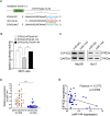
EIF4G2 is overexpressed in HCC and high EIF4G2 expression indicates poor prognosis of HCC patients. a OS analysis in HCC patients with high- or low-expression of EIF4G2 from the Human Protein Atlas database. b Representative images of EIF4G2 expression in 30 paired HCC tissues(T) and adjacent para-carcinoma tissues(N). c Representative images of EIF4G2 IHC staining with different staining scores. Images were presented at 2x magnification (up panel) and 20x magnification (lower panel). d Representative images and analysis of EIF4G2 IHC staining in HCC tissues and adjacent para-carcinoma tissues, images were presented at 2x magnification. e Kaplan–Meier curves analysis of OS in HCC-TMA patients with high- or low-expression of EIF4G2. f Kaplan–Meier curves demonstrating DFS in HCC-TMA patients with high- or low-expression of EIF4G2. g Spearman’s correlation analysis of EIF4G2 expression and PDL1 expression. *p < 0.05
Fig. 2
Fig. 2
EIF4G2 facilitates HCC growth and metastasis in vitro. a HCC cells Hep3B and Huh7 cells were transfected with negative control (NC) or three different si-RNAs of EIF4G2. The knockdown efficiency of EIF4G2 was measured by Western blot. b Functions of EIF4G2 knockdown on HCC cells proliferation were performed by colony formation assay. c RTCA analysis of cells proliferation in Hep3B and Huh7 cells. d Functions of EIF4G2 knockdown on HCC cells migration were determined by scratch experiment. e Effects of EIF4G2 knockdown on HCC cells invasion were detected by transwell invasion assays. *p < 0.05, **p < 0.01, ***p < 0.001
Fig. 3
Fig. 3
EIF4G2 activates the ERK signaling pathway in HCC. a KEGG pathway enrichment analysis of top 20 differentially expressed signaling pathways in EIF4G2 knockdown group compared with ctrl group. b MAPK signaling pathway was analyzed by Western blot. c ERK signaling pathway was tested by Western blot
Fig. 4
Fig. 4
EIF4G2 knockdown suppresses tumorigenesis in vivo. a Fluorescent photos of nude mice bearing HCC tumors. b Representative of HCC tumors from NC and si-EIF4G2 groups. c The tumor growth curves. d HCC tumor weight was measured, and the results were presented as mean ± S.D. e The expression of EIF4G2, ERK1/2 and p-ERK1/2 proteins in mice tumor were measured with Western blot. f Relative signal intensity of EIF4G2 and p-ERK1/2 proteins level of WB. *p < 0.05, ** p < 0.01
Fig. 5
Fig. 5
EIF4G2 is negatively regulated by miR-144. a Putative binding sites between miR-144 and EIF4G2-WT or EIF4G2-MUT 3’UTR position. b Luciferase reporter vectors of EIF4G2-WT or EIF4G2-MUT and miR-144 overexpression vectors were co-transfected into 293 T cells. Relative luciferase activity was assessed 48 h later. (C) Western blot analysis of EIF4G2 level. d The expression of miR-144 in HCC tumor tissues and matched para-cancer tissues. e Pearson association analysis of the miR-144 level and EIF4G2 protein expression. **p < 0.01, ****p < 0.0001 WT: the wild-type vector, MUT: the mutant vector
Fig. 6
Fig. 6
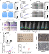
MiR-144 suppresses HCC development in vitro and in vivo. a Functions of overexpression of miR-144 on Hep3B and Huh7 cells were determined by colony formation assay. b RTCA analysis of cells proliferation in two HCC cells (The vertical line represented the time of cell transfection). c Cells migration ability was assessed by wound healing assay. d Cells invasion ability was measured by transwell invasion assay. e Statistical analysis of the invaded cells. f Fluorescent photos of nude mice bearing HCC tumors. g Representative of HCC tumors from NC and miR-144 overexpression groups. h The tumor growth curves. i HCC tumor weight was measured. j Representative images of EIF4G2 IHC staining of mice tumor sections from NC and miR-144 group. * p < 0.05, **p < 0.01
Fig. 7
Fig. 7
Functions of EIF4G2 in HCC are regulated by miR-144. a Western blot analysis of EIF4G2, ERK1/2 and p-ERK1/2 expression. b The migration ability was performed with scratch experiments. c The invasion ability was assessed by transwell invasion assays. **p < 0.01

References

    1. Siegel RL, Miller KD, Jemal A. Cancer statistics, 2018. CA Cancer J Clin. 2018;68(1):7–30. doi: 10.3322/caac.21442. - DOI - PubMed
    1. Bruix J, Reig M, Sherman M. Evidence-based diagnosis, staging, and treatment of patients with hepatocellular carcinoma. Gastroenterology. 2016;150(4):835–853. doi: 10.1053/j.gastro.2015.12.041. - DOI - PubMed
    1. Bhat M, Robichaud N, Hulea L, Sonenberg N, Pelletier J, Topisirovic I. Targeting the translation machinery in cancer. Nat Rev Drug Discov. 2015;14(4):261–278. doi: 10.1038/nrd4505. - DOI - PubMed
    1. Gantenbein N, Bernhart E, Anders I, Golob-Schwarzl N, Krassnig S, Wodlej C, et al. Influence of eukaryotic translation initiation factor 6 on non-small cell lung cancer development and progression. Eur J Cancer. 2018;101:165–180. doi: 10.1016/j.ejca.2018.07.001. - DOI - PubMed
    1. Golob-Schwarzl N, Krassnig S, Toeglhofer AM, Park YN, Gogg-Kamerer M, Vierlinger K, et al. New liver cancer biomarkers: PI3K/AKT/mTOR pathway members and eukaryotic translation initiation factors. Eur J Cancer. 2017;83:56–70. doi: 10.1016/j.ejca.2017.06.003. - DOI - PubMed

MeSH terms