Case Reports
. 2021 Feb 1;14(2):e237401.
doi: 10.1136/bcr-2020-237401.

Multiple thromboembolic events associated with bilateral superior vena cava and anomalous drainage into the left atrium

Affiliations
Case Reports

Multiple thromboembolic events associated with bilateral superior vena cava and anomalous drainage into the left atrium

Maria Elizabeth Karavassilis et al. BMJ Case Rep. .

Abstract

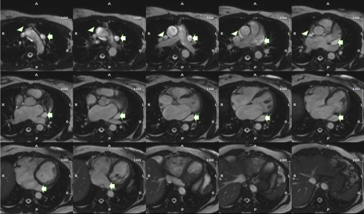
A 49-year-old female patient presented with acute-on-chronic chest pain. She was diagnosed with multiple systemic thromboemboli, including myocardial infarctions, bilateral chronic pulmonary emboli, ischaemic stroke, deep venous thrombosis and superficial thrombophlebitis. She had a background of sickle cell trait. Cardiac magnetic resonance showed bilateral superior vena cava (SVC). The right-sided SVC (RSVC) was joined by the right upper pulmonary vein and drained anomalously into the left atrium. This caused a small volume right to left shunt. The persistent left SVC drained into the right atrium (RA) via a dilated coronary sinus. The overall clinical impression was recurrent paradoxical emboli due to anomalous venous anatomy with a thrombophilia secondary to sickle cell trait. In the normal embryo, the right common cardinal vein develops to become the RSVC, which drains into the RA by term.

Keywords: clinical diagnostic tests; haematology (incl blood transfusion); radiology; radiology (diagnostics); venous thromboembolism.


Conflict of interest statement

Competing interests: None declared.

Figures

Figure 1
Figure 1
Transthoracic echocardiogram (lesion indicated with arrows). (A) Parasternal long axis view: arrowhead pointing to the dilated coronary sinus. (B) Two-chamber view: arrowhead pointing to the dilated coronary sinus. (C) Modified right ventricular inflow view: arrowhead pointing to the ostium of the dilated coronary sinus. (D) Right clavicle view: arrowhead pointing to the right-sided superior vena cava (RSVC). (E) Left clavicle view: arrowhead pointing to the left-sided superior vena cava (LSVC). (F) Left clavicle view: arrowhead pointing to the LSVC.
Figure 2
Figure 2
Cardiac MRI: Late phase after gadolinium contrast showing an area of subendocardial myocardial infarction in the basal inferolateral left ventricle.
Figure 3
Figure 3
Cardiac MRI (lesions indicated with arrows): (A) Four-chamber view: still image from steady state-free precession (SSFP) cine showing a dilated coronary sinus. (B) Two-chamber view: still image from SSFP cine showing a dilated coronary sinus. (C) Three-chamber view: still image from SSFP cine showing a dilated coronary sinus.
Figure 4
Figure 4
Cardiac MRI CT MPR (multiplanar reconstruction) showing the right-sided superior vena cava (SVC) entering the left atrium (arrow), and an anomalous right upper pulmonary vein entering the right-sided SVC (arrowhead).
Figure 5
Figure 5
Cardiac MRI transaxial stack of bright blood steady state-free precession (SSFP) images showing the eft-sided superior vena cava (SVC) draining into dilated coronary sinus (arrow) and right-sided SVC being joined by anomalous right upper pulmonary vein (third panel) and draining into the left atrium (fifth panel) (arrowhead).

References

    1. Connors JM Thrombophilia testing and venous thrombosis. N Engl J Med 2017;377:1177–87. 10.1056/NEJMra1700365 - DOI - PubMed
    1. Windecker S, Stortecky S, Meier B. Paradoxical embolism. J Am Coll Cardiol 2014;64:403–15. 10.1016/j.jacc.2014.04.063 - DOI - PubMed
    1. Saha S, Paoletti D, Robertson M. Persistent left superior vena cava - considerations in fetal, pediatric and adult populations. Australas J Ultrasound Med 2012;15:61–6. 10.1002/j.2205-0140.2012.tb00228.x - DOI - PMC - PubMed
    1. de Leval MR, Ritter DG, McGoon DC, et al. . Anomalous systemic venous connection. surgical considerations. Mayo Clin Proc 1975;50:599–610. - PubMed
    1. Folsom AR, Tang W, Roetker NS, et al. . Prospective study of sickle cell trait and venous thromboembolism incidence. J Thromb Haemost 2015;13:2–9. 10.1111/jth.12787 - DOI - PMC - PubMed

MeSH terms