. 2021 Mar 23;203(8):e00580-20.
doi: 10.1128/JB.00580-20. Print 2021 Mar 23.

Holin-Dependent Secretion of the Large Clostridial Toxin TpeL by Clostridium perfringens

Affiliations

Holin-Dependent Secretion of the Large Clostridial Toxin TpeL by Clostridium perfringens

Angela Saadat et al. J Bacteriol. .

Abstract

Large clostridial toxins (LCTs) are secreted virulence factors found in several species, including Clostridioides difficile, Clostridium perfringens, Paeniclostridium sordellii, and Clostridium novyi LCTs are large toxins that lack a secretion signal sequence, and studies by others have shown that the LCTs of C. difficile, TcdA and TcdB, require a holin-like protein, TcdE, for secretion. The TcdE gene is located on the pathogenicity locus (PaLoc) of C. difficile, and holin-encoding genes are also present in the LCT-encoded PaLocs from P. sordellii and C. perfringens However, the holin (TpeE) associated with the C. perfringens LCT TpeL has no homology and a different membrane topology than TcdE. In addition, TpeE has a membrane topology identical to that of the TatA protein, which is the core of the twin-arginine translocation (Tat) secretion system. To determine if TpeE was necessary and sufficient to secrete TpeL, the genes from a type C strain of C. perfringens were expressed in a type A strain of C. perfringens, HN13, and secretion was measured using Western blot methods. We found that TpeE was required for TpeL secretion and that secretion was not due to cell lysis. Mutant forms of TpeE lacking an amphipathic helix and a charged C-terminal domain failed to secrete TpeL, and mutations that deleted conserved LCT domains in TpeL indicated that only the full-length protein could be secreted. In summary, we have identified a novel family of holin-like proteins that can function, in some cases, as a system of protein secretion for proteins that need to fold in the cytoplasm.IMPORTANCE Little is known about the mechanism by which LCTs are secreted. Since LCTs are major virulence factors in clostridial pathogens, we wanted to define the mechanism by which an LCT in C. perfringens, TpeL, is secreted by a protein (TpeE) lacking homology to previously described secretion-associated holins. We discovered that TpeE is a member of a widely dispersed class of holin proteins, and TpeE is necessary for the secretion of TpeL. TpeE bears a high degree of similarity in membrane topology to TatA proteins, which form the pore through which Tat secretion substrates pass through the cytoplasmic membrane. Thus, the TpeE-TpeL secretion system may be a model for understanding not only holin-dependent secretion but also how TatA proteins function in the secretion process.

Keywords: Clostridium; molecular genetics; perfringens; secretion systems; toxin.


Figures

FIG 1
FIG 1
Gene synteny, membrane topology, and sequence homology of LCT-associated holin-like proteins in clostridia. (A) Gene organization in LCT PaLocs and a bacteriocin-containing operon. Light blue, LCTs and bacteriocin; yellow, sigma factors; orange, holin-like proteins. Gene lengths are not to scale. (B) Proposed membrane topology of holins and holin-like proteins from panel A and twin-arginine transport (Tat) proteins. TatA and TatE are from E. coli, and TatAd is from B. subtilis. (C) Sequence alignments of holin-like proteins. The proteins were aligned using the ClustalW algorithm in the MegAlign software suite, part of the Lasergene 16 DNA analysis package from DNAStar. Residues that match the TpeE sequence are highlighted in yellow shading. Note that there are 7 residue differences between TpeE and UviB. GenBank sequence accession numbers are as follows: WP_003453321.1 for C. perfringens TpeE, ABG87870.1 for C. perfringens UviB, WP_105463372.1 for E. coli TatE, and QNN28895.1 for B. subtilis TatAd. The PDB ID for E. coli TatA is 2MN7_A.
FIG 2
FIG 2
Schematic diagram showing plasmid constructs used for heterologous expression of tpeE and tpeL in C. perfringens strain HN13. pKRAH1 has a lactose-inducible promoter for regulated expression (31), which is indicated by arrows. ΔHCD, deletion of the high charge density domain at the C terminus; ΔAPH, deletion of the amphipathic helix. TpeLA, TpeLAC, and TpeLACD are constructs with deletions of nested C-terminal domains of full-length TpeL. All constructs have a SacII site at the 5′ end and a BamHI site at the 3′ end of the inserts. Some, indicated with an asterisk, have an additional SalI site inserted between the tpeE and tpeL genes to allow efficient cloning. Each plasmid and its description are also listed in Table 1.
FIG 3
FIG 3
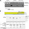
Efficient TpeL secretion is TpeE dependent in C. perfringens. (A) Western blotting using an antibody direct against the His6 tag on TpeL to indicate the location (supernatant or intracellular) of TpeL. pKRAH, empty vector control; pAS11, toxin only; pAS12, toxin and holin; pAS15, holin alone. For the lysis control and loading controls, BCP was identified by fluorescently labeled streptavidin (see Materials and Methods). (B) A C-terminal polyhistidine tag does not significantly affect TpeL expression or secretion in C. perfringens. (Top) Coomassie-stained protein gels demonstrating similar amounts of TpeL in concentrated culture supernatants from strains overexpressing TpeE (no tag) and TpeL with and without a C-terminal His6 tag. Note that there is a protein in the supernatant that is somewhat larger than TpeL; this can be seen clearly in the lane with pKRAH1 and no TpeE (leftmost lane). (Bottom) Anti-His6 immunoblot combined with Ponceau S staining of the PVDF membrane showing similar levels of TpeL in the supernatants (white arrows) whether the His6 tag was present or not. Strains expressing TpeL with and without the His6 tag in the absence of TpeE are shown as secretion negative controls. Whole-cell lysate samples were included to control for differences in TpeL and TpeL-His6 expression. Strains expressing the empty vector and TpeE alone were included to control for effects related to the expression vector and TpeE. Note the increased levels of TpeL in the supernatant when the TpeE protein was also present. The results are representative of data from three independent experiments. (C) Sensitivity of lysis detection by the conjugated streptavidin probe. The image shows a Western blot using an antibody against Tpe-His6 and a fluorescent streptavidin conjugate to detect the cytoplasmic BCP of C. perfringens. When the lysate from the toxin-only strain was added to the filtered supernatant and concentrated, lysis was reliably detected at levels as low as 2% of total cell lysis (see Materials and Methods). +C represents a positive control, using pAS11 to express the toxin, and −C represents a negative control with the empty vector, pKRAH1. The image is representative of results from two separate biological experiments. MW, molecular weight marker.
FIG 4
FIG 4
Expression of tpeE in C. perfringens strain HN13 did not negatively affect the growth rate, while overexpression of tpeL decreased the growth rate. Shown are results for pKRAH1 (vector control), pAS15 (tpeE), pAS11 (tpeL), and pAS12 (tpeE-tpeL). (A) Results after induction with lactose; (B) controls with no lactose added. The means and standard deviations (SD) from five replicate samples are shown. The curves are representative of results from three independent biological replicates.
FIG 5
FIG 5
(A) TpeE localizes to the membrane in C. perfringens. Western blotting was performed using anti-FLAG antibodies to identify the cellular location of TpeE-FLAG. Plasmids pKRAH1 (vector control), pAS13 (tpeE), and pAS15 (tpeE-FLAG) were used. The cytoplasmic loading control consisted of a fluorescent streptavidin conjugate to detect the cytoplasmic BCP of C. perfringens. (B) Detection of tpeE transcripts in C. perfringens using RT-PCR. (Top) Location of primers used for RT-PCR. (Bottom) Agarose gel showing the relative sizes of RT-PCR products and the conditions used for each sample.
FIG 6
FIG 6
TpeE is toxic to and localizes to the membrane and cytoplasm of E. coli. (A) Growth curves with E. coli showing the toxic effects of adding increasing concentrations of the inducer arabinose at the time of inoculation. The means and SD from five replicate samples are shown. The curve is representative of data from three independent biological replicates. (B) Arabinose induction inhibits the growth of colonies containing the tpeE gene (pAS17 and pAS18) on agar plates in E. coli. (C) Western blot showing that TpeE-FLAG was detected in whole-cell lysates, the cytoplasm, and membranes when expressed in E. coli. The cytoplasmic loading controls represent the use of a fluorescent streptavidin conjugate to detect the cytoplasmic biotin carboxyl carrier protein AccB (22.5 kDa) in E. coli. (D) Western blot showing the strains of E. coli in which TpeE appeared in the supernatant after lysis along with the AccB cytoplasmic protein (detected using a fluorescent streptavidin conjugate).
FIG 7
FIG 7
Conserved domains in TpeE are required for secretion of TpeL by C. perfringens. (A) Diagram showing the predicted cellular location of each domain in TpeE. The amphipathic nature of the central alpha-helix is shown using a helical wheel projection of hydrophobic (yellow) and hydrophilic (all other colors) residues. TMD, transmembrane domain. (B) Western blots showing the presence of TpeL-His6 and TpeE-FLAG in whole-cell lysates and supernatants. Note the lack of a TpeL-His6 signal in the supernatants of the strains expressing the TpeE protein with the amphipathic helix or C-terminal domain deleted, even though no cell lysis could be detected with the streptavidin-BCP control.
FIG 8
FIG 8
Truncations of the TpeL toxin lead to loss of secretion. (A) Diagram showing the location of specific domains in the TpeL protein. A, glycosyltransferase domain; C, autocatalytic protease domain; D, delivery domain; B, receptor domain. A table listing the TpeL truncations used and their respective molecular weights is included. (B) Western blot showing the intracellular levels and secretion of full-length and truncated TpeL. An anti-His6 antibody was used to detect each form of the protein. Note the absence of the streptavidin-BCP lysis control signal in the supernatant fractions, indicating that very little lysis had occurred. (C) Growth curves showing that strains expressing truncated TpeL proteins (pAS42, pAS43, and pAS44) do not have defects in their growth in comparison to strains expressing full-length TpeL (pAS11 and pAS45). The means and standard errors of the means (SEM) from five replicate samples are shown. The curves are representative of results from three independent biological replicates.
FIG 9
FIG 9
Model showing a potential mechanism of TpeE-dependent secretion of TpeL. Hydrophobic residues are shown in orange in the TpeL model. The sequence shown is based on the charged zipper mechanism proposed for the Tat secretion system (41), where TatA is a membrane topology model for TpeE. The main features of the model are described in Discussion. The asterisks represent potential salt bridges between charged residues in the amphipathic helix (APH) and high charge density (HCD) domains. The model for the TpeL structure was made using the TcdA structure as a template with the Swiss Model software package. The hydrophobic surface residues were identified using the PyMOL molecular graphics system. TMH, transmembrane helix.

Similar articles

Cited by

References

    1. Navarro MA, McClane BA, Uzal FA. 2018. Mechanisms of action and cell death associated with Clostridium perfringens toxins. Toxins (Basel) 10:212. doi:10.3390/toxins10050212. - DOI - PMC - PubMed
    1. Schirmer J, Aktories K. 2004. Large clostridial cytotoxins: cellular biology of Rho/Ras-glucosylating toxins. Biochim Biophys Acta 1673:66–74. doi:10.1016/j.bbagen.2004.03.014. - DOI - PubMed
    1. Pauillac S, D’Allayer J, Lenormand P, Rousselle JC, Bouvet P, Popoff MR. 2013. Characterization of the enzymatic activity of Clostridium perfringens TpeL. Toxicon 75:136–143. doi:10.1016/j.toxicon.2013.07.003. - DOI - PubMed
    1. Guttenberg G, Hornei S, Jank T, Schwan C, Lu W, Einsle O, Papatheodorou P, Aktories K. 2012. Molecular characteristics of Clostridium perfringens TpeL toxin and consequences of mono-O-GlcNAcylation of Ras in living cells. J Biol Chem 287:24929–24940. doi:10.1074/jbc.M112.347773. - DOI - PMC - PubMed
    1. Nagahama M, Ohkubo A, Oda M, Kobayashi K, Amimoto K, Miyamoto K, Sakurai J. 2011. Clostridium perfringens TpeL glycosylates the Rac and Ras subfamily proteins. Infect Immun 79:905–910. doi:10.1128/IAI.01019-10. - DOI - PMC - PubMed

Publication types

MeSH terms