. 2021 Feb 19;134(4):jcs258356.
doi: 10.1242/jcs.258356.

Importin α phosphorylation promotes TPX2 activation by GM130 to control astral microtubules and spindle orientation

Affiliations

Importin α phosphorylation promotes TPX2 activation by GM130 to control astral microtubules and spindle orientation

Haijing Guo et al. J Cell Sci. .

Abstract

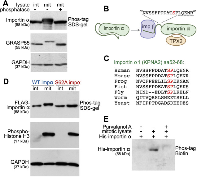
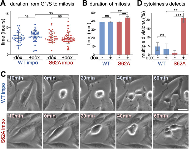
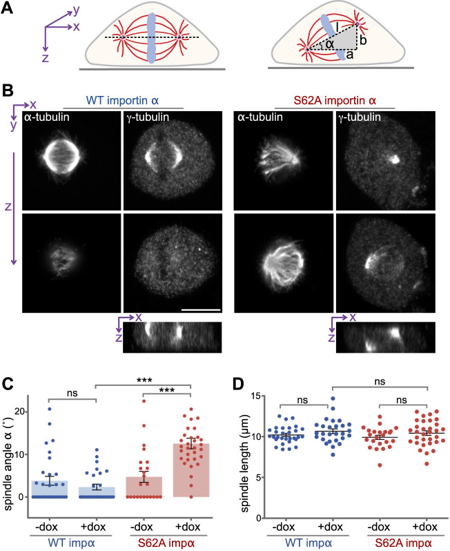
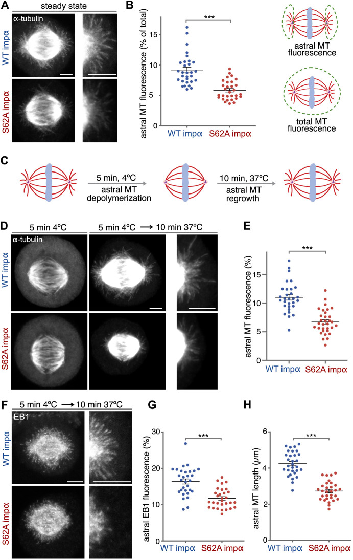
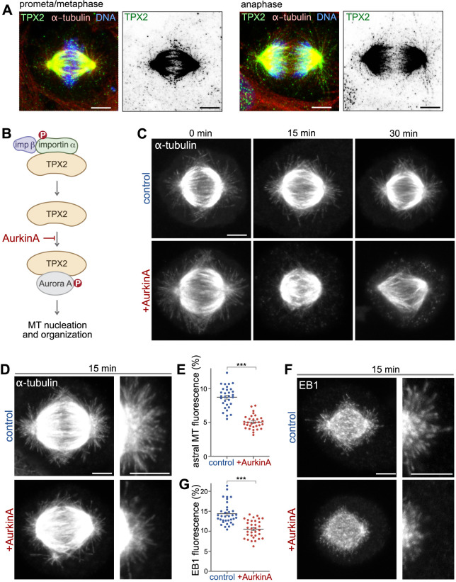
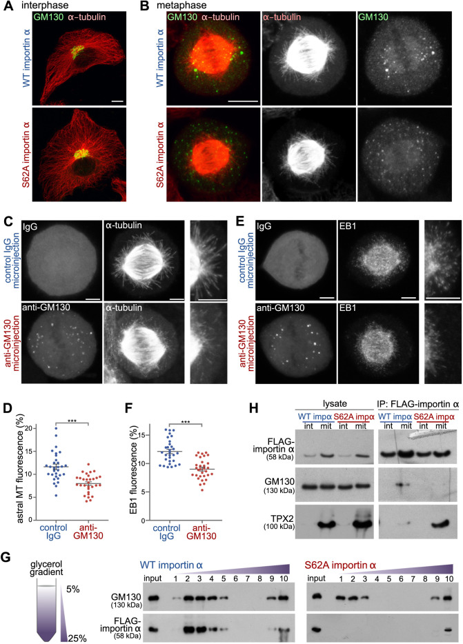
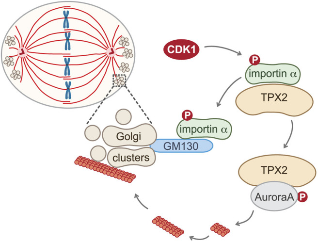
Spindle orientation is important in multiple developmental processes as it determines cell fate and function. The orientation of the spindle depends on the assembly of a proper astral microtubule network. Here, we report that the spindle assembly factor TPX2 regulates astral microtubules. TPX2 in the spindle pole area is activated by GM130 (GOLGA2) on Golgi membranes to promote astral microtubule growth. GM130 relieves TPX2 inhibition by competing for importin α1 (KPNA2) binding. Mitotic phosphorylation of importin α at serine 62 (S62) by CDK1 switches its substrate preference from TPX2 to GM130, thereby enabling competition-based activation. Importin α S62A mutation impedes local TPX2 activation and compromises astral microtubule formation, ultimately resulting in misoriented spindles. Blocking the GM130-importin α-TPX2 pathway impairs astral microtubule growth. Our results reveal a novel role for TPX2 in the organization of astral microtubules. Furthermore, we show that the substrate preference of the important mitotic modulator importin α is regulated by CDK1-mediated phosphorylation.

Keywords: Astral microtubules; CDK1; GM130; Golgi; Importin α; Mitosis; Phosphorylation; Spindle orientation; TPX2.


Conflict of interest statement

Competing interestsThe authors declare no competing or financial interests.

Figures

Fig. 1.
Fig. 1.
Importin α is mitotically phosphorylated at serine 62 by CDK1. (A) Importin α is phosphorylated during mitosis. Cell lysates from interphase (int) and mitotic (mit) HeLa cells, as well as λ phosphatase-treated mitotic lysate, were separated on a 10% SDS gel containing 25 μM Phos-tag acrylamide and immunoblotted for importin α. GRASP55 and GADPH were separated by regular SDS–PAGE. (B) Schematic illustration of the location of the mitotically-phosphorylated peptide identified by mass spectrometry. The identified peptide (residues 52–68) harbors the mitosis-specific phosphorylation site of importin α1 (KPNA2) and is located between the importin β (imp β)-binding domain and the NLS-binding domain. (C) Within the identified peptide sequence, residue serine 62 is conserved among higher eukaryotes. The Ser-Pro sequence matches a CDK1 phosphorylation consensus motif. (D) Importin α (impα) is phosphorylated at serine 62. Interphase or mitotic cell lysates from cells expressing FLAG-tagged WT importin α or importin α S62A were separated on a Phos-tag acrylamide gel and immunoblotted for FLAG. Phospho-histone H3 and GAPDH were separated by SDS–PAGE. (E) Importin α phosphorylation is blocked by the CDK1 inhibitor purvalanol A. Beads coated with recombinant full-length His-tagged importin α were incubated with mitotic HeLa extract in the presence or absence of purvalanol A. The beads were then washed by high-salt buffer (PBS, 1M NaCl), and importin α was eluted by boiling in SDS sample buffer. Phosphorylation was detected by blotting with Phos-tag biotin and streptavidin–HRP. Data in A,D,E are representative of three experiments.
Fig. 2.
Fig. 2.
Importin α S62A induces mitotic defects. (A–D) NRK cells non-induced or induced using doxycycline (dox) to express WT importin α (impα) or importin α S62A were synchronized at the G1-S transition by means of thymidine treatment for 16 h. The cells were then released from the thymidine block and cell cycle progression was monitored by time-lapse phase-contrast microscopy. (A) Importin α expression does not affect mitotic entry. The duration between release from G1-S arrest to mitotic entry is plotted. n>90. ns indicates P=0.075 for WT +dox versus S62A +dox, P=0.211 for WT −dox versus WT +dox and P=0.316 for S62A −dox versus S26A +dox (unpaired, two-tailed Student's t-test). (B) Importin α S62A-expressing cells exhibit prolonged mitotic duration. n>90. **P=0.0065 for WT +dox versus S62A +dox and **P=0.0062 for S62A −dox versus S26A +dox; ns, P=0.93 for WT −dox versus WT +dox (unpaired, two-tailed Student's t-test). (C,D) NRK cells expressing importin α S62A exhibit cytokinesis defects. Still images from a representative time-lapse movie at the indicated time points after mitotic entry (0 min) are shown in C. n>90. ***P=0.0004; **P=0.0017; ns P=0.545 (unpaired, two-tailed Student's t-test). Data are presented as mean±s.d. Scale bar: 20 µm.
Fig. 3.
Fig. 3.
Phosphorylation of importin α controls spindle orientation. (A) Schematic illustration of spindle orientation in tissue culture cells. The axis of a normal spindle is oriented parallel to the coverslip (left), whereas the axis of a misoriented spindle is tilted (right). The angle α of spindle tilt was calculated as: α=arctan (b/a). Spindle length (l) was calculated as the 3D distance between the spindle poles (l2=a2+b2). (B) Importin α S62A causes misorientation of the spindle. NRK cells expressing WT importin α or importin α S62A were arrested at metaphase using MG132 and then dual-stained for α-tubulin and γ-tubulin to label spindle microtubules and the spindle poles, respectively. Z-sections were captured by confocal microscopy. Orthogonal sections through the z-stacks along the x-axis (bottom panels) indicate the relative spindle pole localization on the z-axis. Scale bar: 10 μm. (C) Importin α (impα) S62A results in tilted spindles. The spindle tilt angle α was calculated as shown in A for cells with or without doxycyclin (dox) treatment. n>30. ***P<0.0001; ns, WT −dox versus WT +dox P=0.2478 (Student's t-test). (D) Importin α S62A expression does not alter spindle length. The spindle length l was determined as depicted in A. n>30. ns for WT +dox versus S62A +dox indicates P=0.5676, WT −dox versus WT +dox P=0.185 and S62A −dox versus S62 +dox P=0.196 (Student's t-test). Data are presented as mean±s.e.m.
Fig. 4.
Fig. 4.
Importin α S62A inhibits microtubule growth. (A) Importin α (impα) S62A compromises astral microtubules. NRK cells expressing WT importin α or importin α S62A were arrested at metaphase using MG132 for 1 h and stained for α-tubulin. Maximum intensity projections of confocal z-sections are shown. Enlargements of the astral microtubule area are shown in the panels on the right. (B) Quantitation of the experiment shown in A. Astral microtubule (MT) fluorescence was measured as the percentage of astral microtubule intensity relative to the total microtubule intensity, as shown in the schematic. n>30. ***P<0.0001 (Student's t-test). (C) Schematic illustration of the cold-induced astral microtubule regrowth assay. Astral microtubules were depolymerized by 5 min of cold treatment, and then allowed to regrow by shifting the cells back to 37°C for 10 min. (D) Importin α S62A inhibits astral microtubule regrowth upon cold treatment. NRK cells arrested at metaphase using MG132 were placed on ice for 5 min to depolymerize astral microtubules but not spindle filaments. After 10 min regrowth at 37°C, cells were fixed and stained for α-tubulin. Confocal microscopy images represent maximum intensity projections. Enlargements of the astral microtubule area are shown in the panels on the right. (E) Astral microtubule fluorescence was calculated as in B. n>30. ***P<0.0001 (Student's t-test). (F) Importin α S62A inhibits EB1 localization to the astral area. EB1 was stained following the procedure described in C,D. Maximum intensity projections are shown. Enlargements of the astral microtubule area are shown in the panels on the right. (G) EB1 fluorescence was calculated as for astral microtubule intensity. n>30. ***P<0.0001 (Student's t-test). (H) Importin α S62A shortens astral microtubule length. The lengths of astral microtubules were determined as the distance between the tip of an EB1 signal comet and the spindle pole. For each cell, the average length of the three longest filaments from each pole was used to represent nascent microtubule length. n>30. ***P=0.019 (Student's t-test). Data in B,E,G,H are presented as mean±s.e.m. Scale bars: 5 µm.
Fig. 5.
Fig. 5.
TPX2 is required for astral microtubule formation. (A) Endogenous TPX2 localizes to astral microtubules and spindle microtubules. SV589 cells in prometa/metaphase and anaphase were stained for TPX2 (green), α-tubulin (red) and DNA (blue). Panels on the right show the TPX2 signal in inverted grayscale. (B) AurkinA is a small molecule inhibitor that specifically blocks the interaction of TPX2 with Aurora A kinase (MT, microtubule). (C) AurkinA alters spindle organization. NRK cells arrested at metaphase using MG132 for 1 h were treated with AurkinA for 0, 15 or 30 min and then fixed and stained for α-tubulin. (D) AurkinA compromises astral microtubules. After 15 min of AurkinA treatment, NRK cells were fixed and stained for α-tubulin. Enlargements of the astral microtubule area are shown in the panels on the right. (E) Quantitation of the experiment shown in D. n>30, ***P<0.0001 (Student's t-test). (F) AurkinA inhibits astral microtubule growth. After 15 min of Aurkin A treatment, NRK cells were fixed and stained for EB1. Enlargements of the astral microtubule area are shown in the panels on the right. (G) Quantitation of the experiment shown in F. n>30. ***P<0.0001 (Student's t-test). In E,G, data are presented as mean±s.e.m. Maximum intensity projections of confocal z-sections are shown. Scale bars: 5 µm.
Fig. 6.
Fig. 6.
GM130 regulates astral microtubule growth via phosphorylated importin α. (A) Importin α S62A does not alter Golgi structure or the microtubule network during interphase. Interphase NRK cells expressing WT importin α or importin α S62A were fixed and stained for α-tubulin and the Golgi membrane protein GM130. Maximum intensity projections are shown. Scale bar: 10 µm. (B) Importin α S62A delocalizes mitotic Golgi clusters from the spindle pole area. NRK cells expressing WT importin α or importin α S62A were arrested at metaphase using MG132, and then fixed and stained for GM130 and α-tubulin. Maximum intensity projections are shown. Scale bar: 10 µm. (C,E) Blocking the GM130-importin α interaction inhibits astral microtubule growth and delocalizes Golgi clusters from the spindle poles. NRK cells were arrested at metaphase for 1 h and microinjected with inhibitory antibodies against GM130 or with control IgG. Injected cells were incubated for another 30 min at 37°C, followed by cold treatment for 5 min. They were then allowed to regrow microtubules for 10 min, before fixation and staining for α-tubulin (C), EB1 (E) and injected IgG. Maximum intensity projections are shown. Enlargements of the astral microtubule area are shown in the panels on the right. Scale bars: 5 µm. (D,F) Quantitation of astral microtubule (MT) fluorescence (D) and astral EB1 fluorescence (F) from the experiments shown in C and E, respectively. Data are presented as mean±s.e.m. n>30. ***P<0.0001 (Student's t-test). (G) Importin α S62A does not associate with mitotic Golgi membranes. Post-chromosomal supernatant of mitotic HeLa cells expressing FLAG-tagged WT importin α or importin α S62A was centrifuged through a linear 5–25% glycerol gradient to separate membrane components by size (left). Ten fractions were collected from the top. The membranes from each fraction were collected by centrifugation and then analyzed by western blotting of the indicated proteins (right). (H) Phosphorylation of importin α shifts its binding preference from TPX2 to GM130. Interphase (int) or mitotic (mit) HeLa cells expressing FLAG-tagged WT importin α or importin α S62A were lysed and incubated with anti-FLAG antibody-conjugated beads (IP, immunoprecipitation). Beads were then washed with lysis buffer and analyzed by western blotting for the indicated proteins. Data in G and H are representative of three experiments.
Fig. 7.
Fig. 7.
Model of the TPX2-mediated astral microtubule assembly pathway. Phosphorylation (P) of importin α at S62 by CDK1 switches its substrate preference from TPX2 to GM130. This switch enables GM130 on the mitotic Golgi clusters at the spindle poles to locally sequester importin α from TPX2 through direct competition. TPX2 is thereby relieved from inhibition, allowing it to bind to and activate Aurora A kinase to drive astral microtubule assembly and facilitate spindle orientation.

Similar articles

Cited by

References

    1. Anderson, K., Yang, J., Koretke, K., Nurse, K., Calamari, A., Kirkpatrick, R. B., Patrick, D., Silva, D., Tummino, P. J., Copeland, R. A.et al. (2007). Binding of TPX2 to Aurora A alters substrate and inhibitor interactions. Biochemistry 46, 10287-10295. 10.1021/bi7011355 - DOI - PubMed
    1. Asare, A., Levorse, J. and Fuchs, E (2017). Coupling organelle inheritance with mitosis to balance growth and differentiation. Science 355, eaah4701 10.1126/science.aah4701 - DOI - PMC - PubMed
    1. Barr, F. A. and Gruneberg, U (2017). Organelle inheritance—what players have skin in the game? Science 355, 458-459. 10.1126/science.aam6381 - DOI - PubMed
    1. Baudoin, N. C. and Cimini, D (2018). A guide to classifying mitotic stages and mitotic defects in fixed cells. Chromosoma 127, 215-227. 10.1007/s00412-018-0660-2 - DOI - PubMed
    1. Bayliss, R., Sardon, T., Vernos, I. and Conti, E (2003). Structural basis of Aurora-A activation by TPX2 at the mitotic spindle. Mol. Cell 12, 851-862. 10.1016/S1097-2765(03)00392-7 - DOI - PubMed

Publication types

MeSH terms