Review
. 2021 Jul;18(7):465-478.
doi: 10.1038/s41569-020-00503-2. Epub 2021 Feb 1.

Artificial intelligence-enhanced electrocardiography in cardiovascular disease management

Affiliations
Review

Artificial intelligence-enhanced electrocardiography in cardiovascular disease management

Konstantinos C Siontis et al. Nat Rev Cardiol. 2021 Jul.

Abstract

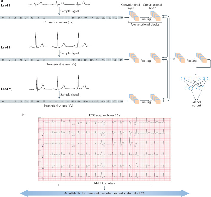
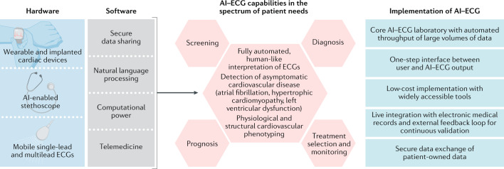
The application of artificial intelligence (AI) to the electrocardiogram (ECG), a ubiquitous and standardized test, is an example of the ongoing transformative effect of AI on cardiovascular medicine. Although the ECG has long offered valuable insights into cardiac and non-cardiac health and disease, its interpretation requires considerable human expertise. Advanced AI methods, such as deep-learning convolutional neural networks, have enabled rapid, human-like interpretation of the ECG, while signals and patterns largely unrecognizable to human interpreters can be detected by multilayer AI networks with precision, making the ECG a powerful, non-invasive biomarker. Large sets of digital ECGs linked to rich clinical data have been used to develop AI models for the detection of left ventricular dysfunction, silent (previously undocumented and asymptomatic) atrial fibrillation and hypertrophic cardiomyopathy, as well as the determination of a person's age, sex and race, among other phenotypes. The clinical and population-level implications of AI-based ECG phenotyping continue to emerge, particularly with the rapid rise in the availability of mobile and wearable ECG technologies. In this Review, we summarize the current and future state of the AI-enhanced ECG in the detection of cardiovascular disease in at-risk populations, discuss its implications for clinical decision-making in patients with cardiovascular disease and critically appraise potential limitations and unknowns.


Conflict of interest statement

P.A.N., Z.I.A., P.A.F. and the Mayo Clinic have filed patents on several AI–ECG algorithms and could receive financial benefit from the use of this technology. At no point will P.A.N., Z.I.A., P.A.F. or the Mayo Clinic benefit financially from its use for the care of patients at the Mayo Clinic.

Figures

Fig. 1
Fig. 1. Development of a convolutional neural network using the 12-lead ECG and application to detect silent atrial fibrillation.
a | The analogue electrocardiogram (ECG) signal is converted to a digital recording, resulting in a list of numerical values corresponding to the amplitude of the signal. (The numerical values depicted are arbitrary and shown for illustrative purposes only.) These numerical values are then convolved with the network weights within each lead and across leads, feeding sequential layers of convolutions until the final model output is reached. b | With the use of a trained, deep-learning artificial intelligence-enhanced ECG (AI–EGG) model, a one-off, standard, 12-lead, sinus-rhythm ECG can become a surrogate for prolonged rhythm monitoring for the detection of silent atrial fibrillation. Part a adapted with permission from ref., Elsevier.
Fig. 2
Fig. 2. The AI–ECG to detect HCM.
Use of an artificial intelligence-enhanced electrocardiogram (AI–ECG) model to detect obstructive hypertrophic cardiomyopathy (HCM) in a woman aged 21 years before (part a) and after (part b) septal myectomy. Adapted with permission from ref., Elsevier.
Fig. 3
Fig. 3. Framework for AI–ECG applications in clinical practice.
Current, versatile electrocardiogram (ECG)-recording technologies (wearable and implantable devices, smartwatches and e-stethoscopes) coupled with the ability to store, transfer, process and analyse large amounts of digital data are increasingly allowing the deployment of artificial intelligence (AI)-powered tools in the clinical arena, addressing the spectrum of patient needs. The science of AI-enhanced ECG (AI–ECG) implementation, including the interface between patients and the AI–ECG output, integration of AI–ECG tools with electronic health records, patient privacy, and cost and reimbursement implications, is in its infancy and continues to evolve.
Fig. 4
Fig. 4. AI–ECG Dashboard linked from within the electronic health record for point-of-care application.
a | A patient with embolic stroke of undetermined source had an increased probability of silent atrial fibrillation (AF; red data points) that predated the clinical documentation of atrial flutter. b | A patient with a history of heart transplantation in 2005 who experienced graft rejection with left ventricular systolic dysfunction in 2020. At that point, the artificial intelligence-enhanced electrocardiogram (AI–ECG) reported a high probability of low ejection fraction (EF), correlating with the graft rejection. AV, atrioventricular; HCM, hypertrophic cardiomyopathy.

References

    1. Krizhevsky A, Sustskever I, Hinton GE. ImageNet classification with deep convolutional neural networks. Adv. Neural Inf. Process. Syst. 2012;25:1106–1114.
    1. Rudin C. Stop explaining black box machine learning models for high stakes decisions and use interpretable models instead. Nat. Mach. Intell. 2019;1:206–215. doi: 10.1038/s42256-019-0048-x. - DOI - PMC - PubMed
    1. Gottesman O, et al. Guidelines for reinforcement learning in healthcare. Nat. Med. 2019;25:16–18. doi: 10.1038/s41591-018-0310-5. - DOI - PubMed
    1. Pipberger HV, Freis ED, Taback L, Mason HL. Preparation of electrocardiographic data for analysis by digital electronic computer. Circulation. 1960;21:413–418. doi: 10.1161/01.CIR.21.3.413. - DOI - PubMed
    1. Caceres CA, Rikli AE. The digital computer as an aid in the diagnosis of cardiovascular disease. Trans. NY Acad. Sci. 1961;23:240–245. doi: 10.1111/j.2164-0947.1961.tb03119.x. - DOI - PubMed

MeSH terms