Review
. 2021 Mar;24(3):297-311.
doi: 10.1038/s41593-020-00778-1. Epub 2021 Feb 1.

Gene-based therapies for neurodegenerative diseases

Affiliations
Review

Gene-based therapies for neurodegenerative diseases

Jichao Sun et al. Nat Neurosci. 2021 Mar.

Abstract

Gene therapy is making a comeback. With its twin promise of targeting disease etiology and 'long-term correction', gene-based therapies (defined here as all forms of genome manipulation) are particularly appealing for neurodegenerative diseases, for which conventional pharmacologic approaches have been largely disappointing. The recent success of a viral-vector-based gene therapy in spinal muscular atrophy-promoting survival and motor function with a single intravenous injection-offers a paradigm for such therapeutic intervention and a platform to build on. Although challenges remain, the newfound optimism largely stems from advances in the development of viral vectors that can diffusely deliver genes throughout the CNS, as well as genome-engineering tools that can manipulate disease pathways in ways that were previously impossible. Surely spinal muscular atrophy cannot be the only neurodegenerative disease amenable to gene therapy, and one can imagine a future in which the toolkit of a clinician will include gene-based therapeutics. The goal of this Review is to highlight advances in the development and application of gene-based therapies for neurodegenerative diseases and offer a prospective look into this emerging arena.


Conflict of interest statement

Author disclosures: J. Sun and S. Roy have applied for patents related to gene editing in Alzheimer’s disease (US Patent App. 16251970). S. Roy is also scientific founder of, advisor to, and owns equity in CRISPRAlz.

Figures

Figure 1:
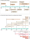
Figure 1:. Timeline of marquee events in the gene therapy field.
a) Milestones in the development of gene therapy tools. b) Timeline of key clinical trials in gene-based therapies for neurodegenerative diseases.
Figure 2.
Figure 2.. Mechanisms of genome editing by engineered nucleases.
a) Double-strand breaks (DSBs, arrowhead) in genomic DNA are repaired by non-homologous end joining (NHEJ), that introduces indel mutations (red dash lines), typically leading to a change of the reading frame, pre-mature termination codon (PTC), and disrupted gene function. b) Alternately, in the presence of donor template, homology-directed repair (HDR) mechanisms lead to precise insertion or modification of DNA (green lines). c) Zinc finger nucleases (ZFNs) are composed of three to six zinc finger domains (color circles), each recognizing three nucleotides. The zinc finger domains on opposite strands of DNA bring two Fok1 endonuclease domains together, inducing Fok1 dimerization and DSB. d) Transcription activator-like effector nucleases (TALENs) have 16-20 TALE monomers, each recognizing a single nucleotide. A pair of TALENs induces Fok1 dimerization and DSB. e) The CRISPR-Cas9 system contains a synthetic guide RNA and the nuclease Cas9. The guide RNA has two domains – a programmable crRNA sequence recognizing the genomic target, and a tracrRNA sequence for Cas9-binding. f) Location of PTCs determine transcription fate. Only PTCs residing >50-55 nt upstream of the last exon-exon junction will lead to non-sense mediated decay (NMD) and degradation of the mRNA. The last exon is exempt from NMD, and PTCs in this “NMD-insensitive region” should lead to truncations (see article for details).
Figure 3.
Figure 3.. Non-canonical CRISPR tools.
a,b) dCas9, a nuclease-deactivated Cas9, is fused to a transcriptional inhibitor (a) or activator (b) to inhibit or boost transcription, respectively. c, d) The dCas9 or nCas9 (a Cas9 nickase) is fused to an adenine (c) or cytosine (d) deaminase that changes A:T base-pairs to G:C or C:G base-pairs to T:A, respectively. e) The nCas9 is fused to a reverse transcriptase that copies the part of prime-editing guide RNA (pegRNA) sequence into the target site (red lines).
Figure 4.
Figure 4.. Mechanisms of antisense oligonucleotides (ASOs).
Normal RNA splicing: The newly synthesized precursor messenger RNA (pre-mRNA) becomes a mature mRNA by RNA-splicing – a process where introns (thin lines) are removed and exons (colored boxes) are ligated. Splice inclusion: ASOs bind to and block the activity of splicing enhancers on the pre-mRNA, leading to the inclusion of an exon that would normally be excluded; generating a modified protein with additional peptide sequences. Splice exclusion: ASOs bind to the exon-intron splicing junction on the pre-mRNA to skip an exon, leading to a truncated protein. mRNA knockdown: ASOs bind to mRNAs and activate RNase H, resulting in mRNA-cleavage and degradation. Translation inhibitor: ASOs bind to the 5’ UTR or coding regions of the mRNA and block translation.
Figure 5.
Figure 5.. ASO and CRISPR-based editing strategies to modulate APP-cleavage products.
a)Left: Schematic showing the transmembrane domain and C-terminus of APP, with corresponding exons (small arrows with numbers denote amino acids). Right: The ASO strategy leads to “skipping” of APP exon 17, leading to a protein that lacks the γ-secretase cleavage site and a portion of the transmembrane domain, and consequently, less Aβ secretion. Sun et al. used a NHEJ-based CRISPR-Cas9 strategy to introduce PTCs in the last exon (exon 18) of APP. Since transcriptional rules dictate that the last exon is exempt from NMD, this method does not lead to mRNA decay, but effectively truncates the last ~ 36 amino acids of APP that includes an endocytic motif triggering APP β-cleavage (see for more details). b-d) Data from editing of APP C-terminus with CRISPR showing attenuation of APP β-cleavage and upregulation of protective α-cleavage (figures reproduced from, shared under a Creative Commons Attribution 4.0 International Licence). b) Human iPSC-derived neurons (WT or isogenic APPV717I knock-in) were transduced with lentiviral vectors carrying APP-gRNA/Cas9 to edit the C-terminus of APP and immunoblotted with C- and N-terminus antibodies. Note selective attenuation of APP signal with C-terminus antibody Y188 after CRISPR-editing. c) Immunoblotting of secreted sAPPα from the media of iPSC-neurons, treated as mentioned in (b). Note increased sAPPα in edited samples, indicating upregulation of protective APP α-cleavage. d) ELISA of secreted Aβ from media, treated as mentioned in (b). Note decreased Aβ in CRISPR-treated samples.

References

    1. Somanathan S, Calcedo R & Wilson JM Adenovirus-Antibody Complexes Contributed to Lethal Systemic Inflammation in a Gene Therapy Trial. Mol Ther 28, 784–793, doi:10.1016/j.ymthe.2020.01.006 (2020). - DOI - PMC - PubMed
    1. Mendell JR et al.Single-Dose Gene-Replacement Therapy for Spinal Muscular Atrophy. N Engl J Med 377, 1713–1722, doi:10.1056/NEJMoa1706198 (2017). - DOI - PubMed
    1. Keeler CE Gene therapy. J Hered 38, 294–298 (1947). - PubMed
    1. Logovinsky V et al.Safety and tolerability of BAN2401--a clinical study in Alzheimer’s disease with a protofibril selective Abeta antibody. Alzheimers Res Ther 8, 14, doi:10.1186/s13195-016-0181-2 (2016). - DOI - PMC - PubMed
    1. Hudry E & Vandenberghe LH Therapeutic AAV Gene Transfer to the Nervous System: A Clinical Reality. Neuron 102, 263, doi:10.1016/j.neuron.2019.03.020 (2019). - DOI - PubMed

Publication types