. 2021 Jan 27;7(1):156-163.
doi: 10.1021/acscentsci.0c01309. Epub 2020 Dec 18.

De Novo Discovery of High-Affinity Peptide Binders for the SARS-CoV-2 Spike Protein

Affiliations

De Novo Discovery of High-Affinity Peptide Binders for the SARS-CoV-2 Spike Protein

Sebastian Pomplun et al. ACS Cent Sci. .

Abstract

The β-coronavirus SARS-CoV-2 has caused a global pandemic. Affinity reagents targeting the SARS-CoV-2 spike protein are of interest for the development of therapeutics and diagnostics. We used affinity selection-mass spectrometry for the rapid discovery of synthetic high-affinity peptide binders for the receptor binding domain (RBD) of the SARS-CoV-2 spike protein. From library screening with 800 million synthetic peptides, we identified three sequences with nanomolar affinities (dissociation constants K d = 80-970 nM) for RBD and selectivity over human serum proteins. Nanomolar RBD concentrations in a biological matrix could be detected using the biotinylated lead peptide in ELISA format. These peptides do not compete for ACE2 binding, and their site of interaction on the SARS-CoV-2-spike-RBD might be unrelated to the ACE2 binding site, making them potential orthogonal reagents for sandwich immunoassays. These findings serve as a starting point for the development of SARS-CoV-2 diagnostics or conjugates for virus-directed delivery of therapeutics.


Conflict of interest statement

The authors declare the following competing financial interest(s): B.L.P. is a cofounder of Amide Technologies and Resolute Bio. Both companies focus on the development of protein and peptide therapeutics.

Figures

Figure 1
Figure 1
SARS-CoV-2-spike-RBD binding peptides with nanomolar affinity were identified by affinity selection–mass spectrometry. (A) Schematic representation of the AS–MS workflow and enriched sequences. In brief, biotinylated SARS-CoV-2-spike-RBD was immobilized on magnetic streptavidin beads and then incubated with peptide libraries. Unbound members were removed by washing. Peptides bound to SARS-CoV-2-spike-RBD were eluted and analyzed by nanoLC–MS/MS. (B) BLI curves for association/dissociation of peptides 14 to SARS-CoV-2-spike-RBD (in kinetic buffer: 1× PBS, pH = 7.2, 0.1% bovine serum albumin, 0.02% Tween-20). While peptide 4 had higher affinity, peptide 1, compared to 2 and 4, had the best solubility and was used for all further investigations. Peptides 2 and 4 precipitated from solution within hours at concentrations greater than 10 μM. Kinetic binding results are reported in Table S1. (C) BLI curves for 1 (blue line) and scrambled analogues of 1 (light and dark gray lines, respectively; sc1, GSVKRWLTYVKNFK; and sc2, RFYVTKGWSNKVLK). (D) Self-competition analysis (BLI association) of 1 to SARS-CoV-2-spike-RBD: peptide 1-biotin immobilized on BLI tips was dipped into solutions containing SARS-CoV-2-spike-RBD and 1 ([RBD] = 500 nM; [1] = 0–16 μM). Increasing the concentration of 1 in solution causes less free RBD available in solution (due to RBD-1 complex formation) and results in a concentration-dependent decrease in BLI response.
Figure 2
Figure 2
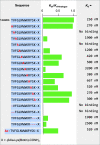
Alanine scanning and sequence truncations of 1 reveal binding hotspots. Binding to SARS-CoV-2-spike-RBD of alanine mutants and truncated peptides was measured by BLI as detailed in Figure 1. Ratios between binding of original sequence 1 and each mutant, respectively, are shown as green bars. Individual steady state Kd values are shown in the right column. Kinetic binding results are reported in Table S1.
Figure 3
Figure 3
Peptide 1 binds to SARS-CoV-1-spike-RBD and MERS-CoV-spike-RBD. (A) The binding of peptide 1-biotin to SARS-CoV-1-spike-RBD and MERS-CoV-spike-RBD was determined by BLI. (B) A structural overlay of SARS-CoV-2-spike-RBD and MERS-CoV-spike-RBD was performed with the software PyMol (using PDB structures 6vw1 and 6c6z). Regions with the homology of the secondary structure between the two proteins are colored in red. The homologous regions were manually analyzed to identify positions with identical residues in both proteins: blue. Since peptide 1-biotin binds to both with comparable affinity, the binding site could potentially be in a region with high homology.
Figure 4
Figure 4
SARS-CoV-2-spike-RBD can be selectively enriched from human serum proteins. (A) Schematic representation of the pull-down of SARS-CoV-2-spike-RBD from human serum. (B) Workflow: spike RBD was added to human serum (RBD: 0.27 mg/mL, 10% human serum, 1× PBS), and the mix was incubated with magnetic beads (MyOne Dynabeads) displaying peptide 1-biotin (1 h, 4 °C). The supernatant (containing nonbinding proteins) was removed, and the beads were washed with 1× PBS (3 × 1 mL). Bound proteins were eluted with 6 M urea (elution 1:50 μL, 30 s; elution 2:50 μL, 120 s) and analyzed by SDS PAGE (C). The gel shows (from left to right) (1) molecular weight ladder; (2) purified SARS-CoV-2-spike-RBD (1 μg); (3) human serum mixed with SARS-CoV-2-spike-RBD; (4) elution 1 (30 μL of elution sample 1); and (5) elution 2 (30 μL of elution sample 2). The analysis was performed using BoltTM 4–12% Bis-Tris Plus gels (10-wells), 165 V for 36 min, utilizing prestained Invitrogen SeeBlueTM Plus2 molecular weight standard with BoltTM LDS sample buffer (4×).
Figure 5
Figure 5
Picomolar SARS-CoV-2-spike-RBD quantities were detected by ELISA. (A) Schematic representation of the ELISA assay. (B) Workflow: Serial dilutions (100 nM to 100 fM) of RBD mixed with fetal bovine serum (FBS) were immobilized on an ELISA plate. The plate was incubated with peptide 1-biotin, followed by streptavidin-HRP and TMB substrate. (C) ELISA absorbance readout at 450 nM as a function of RBD concentrations. Measurements were performed in technical triplicates (n = 3), and statistical significance was calculated with the unpaired t test. RBD 100 nM vs no RBD, p = 0.0012 (**); RBD 100 pM vs no RBD, p = 0.018 (*).

References

    1. World Health Organization . Coronavirus Disease (COVID-19) Weekly Epidemiological Update for August 31st 2020; WHO, 2020.
    1. Webb Hooper M.; Nápoles A. M.; Pérez-Stable E. J. COVID-19 and Racial/Ethnic Disparities. JAMA 2020, 323 (24), 2466.10.1001/jama.2020.8598. - DOI - PMC - PubMed
    1. Tromberg B. J.; Schwetz T. A.; Pérez-Stable E. J.; Hodes R. J.; Woychik R. P.; Bright R. A.; Fleurence R. L.; Collins F. S. Rapid Scaling Up of Covid-19 Diagnostic Testing in the United States — The NIH RADx Initiative. N. Engl. J. Med. 2020, 383 (11), 1071–1077. 10.1056/NEJMsr2022263. - DOI - PMC - PubMed
    1. Carter L. J.; Garner L. V.; Smoot J. W.; Li Y.; Zhou Q.; Saveson C. J.; Sasso J. M.; Gregg A. C.; Soares D. J.; Beskid T. R.; Jervey S. R.; Liu C. Assay Techniques and Test Development for COVID-19 Diagnosis. ACS Cent. Sci. 2020, 6 (5), 591–605. 10.1021/acscentsci.0c00501. - DOI - PMC - PubMed
    1. Zou L.; Ruan F.; Huang M.; Liang L.; Huang H.; Hong Z.; Yu J.; Kang M.; Song Y.; Xia J.; Guo Q.; Song T.; He J.; Yen H.-L.; Peiris M.; Wu J. SARS-CoV-2 Viral Load in Upper Respiratory Specimens of Infected Patients. N. Engl. J. Med. 2020, 382 (12), 1177–1179. 10.1056/NEJMc2001737. - DOI - PMC - PubMed