. 2021 Mar;17(1):9-23.
doi: 10.1007/s11302-021-09763-0. Epub 2021 Feb 1.

That was then, this is now: the development of our knowledge and understanding of P2 receptor subtypes

Affiliations

That was then, this is now: the development of our knowledge and understanding of P2 receptor subtypes

Charles Kennedy. Purinergic Signal. 2021 Mar.

Abstract

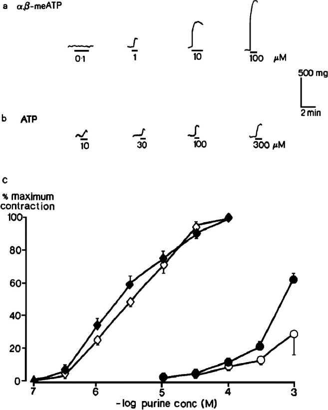
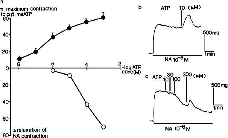
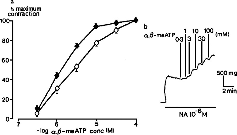
P2 receptors are present in virtually all tissues and cell types in the human body, and they mediate the physiological and pharmacological actions of extracellular purine and pyrimidine nucleotides. They were first characterised and named by Geoff Burnstock in 1978, then subdivided into P2X and P2Y purinoceptors in 1985 on the basis of pharmacological criteria in functional studies on native receptors. Molecular cloning of receptors in the 1990s revealed P2X receptors to comprise seven different subunits that interact to produce functional homo- and heterotrimeric ligand-gated cation channels. A family of eight P2Y G protein-coupled receptors were also cloned, which can form homo- and heterodimers. Deep insight into the molecular mechanisms of agonist and antagonist action has been provided by more recent determination of the tertiary and quaternary structures of several P2X and P2Y receptor subtypes. Agonists and antagonists that are highly selective for individual subtypes are now available and some are in clinical use. This has all come about because of the intelligence, insight and drive of the force of nature that was Geoff Burnstock.

Keywords: G protein–coupled receptor; Heterodimer; Ligand-gated cation channel; P2 receptors; P2X receptors; P2Y receptors.


Conflict of interest statement

The author declares that he has no conflict of interest.

Figures

Fig. 1
Fig. 1
Geoff Burnstock. Geoff Burnstock is seen at his desk in his office at UCL in 1997 pointing at the author’s PhD thesis
Fig. 2
Fig. 2
Contractions of rat isolated femoral artery. Contractions evoked by (a) α,β-methyleneATP (10−7–10−4 M) and (b) ATP (10−5−3 × 10−4 M) at resting tone when endothelium was intact are shown; (c) log concentration-response curves for contractions evoked by α,β-methyleneATP (10−7–10−4 M) (◊,♦) and ATP (10−5–10−3 M) (○,●) at resting tone when endothelium was intact (open symbols) or removed (closed symbols) (n = 6) are shown. Vertical bars show sem. Reproduced from [25] (Kennedy et al., 1985, with permission from Elsevier)
Fig. 3
Fig. 3
Two types of response to ATP in rat isolated femoral artery. a The effects of ATP (10−6–3 × 10−4 M) in tissues precontracted by 10−6 M noradrenaline (NA) when endothelium was intact (o) or removed (●) (n = 4) are shown. Vertical bars represent sem. b Endothelium removed, contraction to 10−5 M ATP. c Endothelium intact, relaxations to ATP (10−3–3 × 10−4 M). Reproduced from [25] (Kennedy et al., 1985, with permission from Elsevier)
Fig. 4
Fig. 4
One type of response to α,β-methyleneATP in rat isolated femoral artery. a The effect of α,β-methyleneATP (3 × 10−7–10−4 M) in tissues precontracted by 10−6 M noradrenaline (NA) when endothelium was intact (o) or removed (●) (n = 6) are shown. Vertical bars show sem. b Contractions evoked by α,β-methyleneATP when the endothelium was intact are shown. Reproduced from [25] (Kennedy et al., 1985, with permission from Elsevier)
Fig. 5
Fig. 5
Relaxations of rat isolated portal vein longitudinal muscle. a Relaxations of precontracted tissues induced by 2-methyithioATP (10−4 M) and ATP (10−4 M) in the same preparation are shown. b The mean peak amplitude of relaxations evoked by 2-methylthioATP (10−6–10−4 M) (Δ) and ATP (10−5-3 × 10−4 M) (O) (n = 6) are shown. Vertical bars show sem. Adapted from [26] (Kennedy and Burnstock, 1985, with permission from Elsevier)
Fig. 6
Fig. 6
Subunit fold and closed, resting conformation of zebrafish P2X4.1 receptor. a The ΔzfP2X4 subunit has a dolphin-like shape. Alpha helices (TM1–2 and α2–5), beta strands (β1–14), disulphide bonds (SS1–5) and attached glycans (g2 and 4) are indicated. b Left-hand panel: a sagittal section reveals a closed conformation of the pore and shows that the gate is located about halfway across the membrane bilayer. Three vestibules (upper, central and extracellular vestibules) are located on the molecular 3-fold axis, with the extracellular vestibule connected to the bulk solution through a fenestration (orange arrow). Right-hand panel: pore lining surface calculated by the Hole49 program. Each colour represents a different radius range measured from the receptor centre (red: <1.15 Å, green: 1.15–2.3 Å, and purple: >2.3 Å). (Reproduced from [42] Kawate et al., 2009, with permission from the Nature Publishing Group)
Fig. 7
Fig. 7
The P2Y1R in complex with MRS2500 and BPTU. Side views of the P2Y1R in complex with (a) MRS2500 and (b) BPTU are shown. The receptor is shown as blue (a) and orange (b) cartoon representation. MRS2500 and BPTU are shown in sphere representation with magenta and green carbons, respectively. The membrane boundaries (brown) are adapted from the OPM database31 with P2Y12R (PDB ID: 4NTJ) as a model (reproduced from [72] Zhang et al., 2015, with permission from the Nature Publishing Group)

References

    1. Lohmann K. Über die pyrophosphate fraktion im muskel. Naturwiss. 1929;17:624–625. doi: 10.1007/BF01506215. - DOI
    1. Fiske CH, SubbaRow Y. Phosphorus compounds of muscle and liver. Science. 1929;70:381–382. doi: 10.1126/science.70.1816.381-a. - DOI - PubMed
    1. Drury AN, Szent-Györgi A. The physiological activity of adenine compounds with special reference to their action upon the mammalian heart. J Physiol. 1929;68:213–237. doi: 10.1113/jphysiol.1929.sp002608. - DOI - PMC - PubMed
    1. Burnstock G. A basis for distinguishing two types of purinergic receptor. In: Straub RW, editor. Cell membrane receptors for drugs and hormones: a multidisciplinary approach. New York: Raven Press; 1978. pp. 107–118.
    1. Van Calker D, Muller M, Hamprecht B. Adenosine regulates via two different types of receptors, the accumulation of cyclic AMP in cultured brain cells. J Neurosci. 1979;33:999–1005. doi: 10.1111/j.1471-4159.1979.tb05236.x. - DOI - PubMed

Substances

LinkOut - more resources