Review
. 2021 Feb 1;131(3):e144918.
doi: 10.1172/JCI144918.

Bite of the wolf: innate immune responses propagate autoimmunity in lupus

Review

Bite of the wolf: innate immune responses propagate autoimmunity in lupus

Sarthak Gupta et al. J Clin Invest. .

Abstract

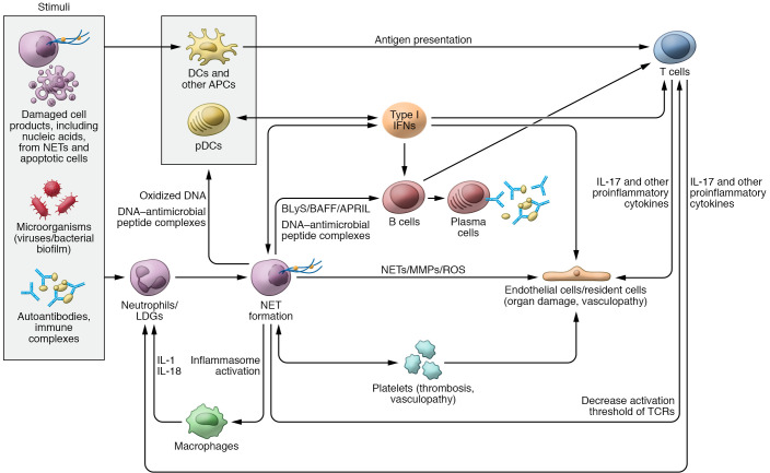
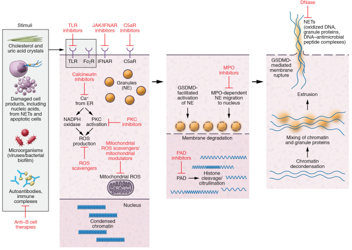
The etiopathogenesis of systemic lupus erythematosus (SLE), a clinically heterogeneous multisystemic syndrome that derives its name from the initial characterization of facial lesions that resemble the bite of a wolf, is considered a complex, multifactorial interplay between underlying genetic susceptibility factors and the environment. Prominent pathogenic factors include the induction of aberrant cell death pathways coupled with defective cell death clearance mechanisms that promote excessive externalization of modified cellular and nuclear debris with subsequent loss of tolerance to a wide variety of autoantigens and innate and adaptive immune dysregulation. While abnormalities in adaptive immunity are well recognized and are key to the pathogenesis of SLE, recent findings have emphasized fundamental roles of the innate immune system in the initiation and propagation of autoimmunity and the development of organ damage in this disease. This Review focuses on recent discoveries regarding the role of components of the innate immune system, specifically neutrophils and interferons, in promoting various aspects of lupus pathogenesis, with potential implications for novel therapeutic strategies.


Conflict of interest statement

Conflict of interest: The NIAMS has collaborative research agreements with MedImmune/AstraZeneca, Pfizer, and Bristol Myers Squibb. MJK is on the scientific advisory boards of Neutrolis, Inc. and Cytrill.

Figures

Figure 1
Figure 1. Role of neutrophils, NETs, and IFNs in SLE pathogenesis.
Various stimuli can trigger neutrophils to undergo neutrophil extracellular trap (NET) formation. NETs, in turn, can externalize self-antigens, including oxidized DNA and/or DNA–antimicrobial peptide complexes that can be presented to antigen-presenting cells (APCs) and activate plasmacytoid DCs (pDCs) to synthesize type I IFNs. NETs have the ability to activate the NLRP3 inflammasome in macrophages, resulting in increased release of IL-1 and IL-18, which further prime neutrophils to undergo NET formation and perpetuate tissue damage. Different exogenous and endogenous stimuli can promote type I IFN generation. The synthesis of type I IFNs further modulates other APCs, tissue-resident cells, and T and B cell functions. NET products and IFNs modulate T cell responses and can also activate B cells to undergo class switching and secrete autoantibodies against a wide range of self-antigens. DNA–antimicrobial peptide complexes (like LL37-DNA) released from NETs have the ability to directly activate B cells via TLR9 and promote autoantibody generation. NETs directly stimulate T cells by decreasing their activation threshold via Zap70-mediated phosphorylation of the T cell receptor (TCR). Activated T cells release IL-17 and other proinflammatory cytokines that can result in endothelial cell damage as well prime neutrophils to undergo further NET formation and migrate to inflamed tissues. NETs and IFNs can promote direct tissue damage and vascular inflammation through their effect on endothelial cells and platelets. APRIL, a proliferation-inducing ligand; BAFF, B cell activating factor; BLyS, B lymphocyte stimulator; LDG, low-density granulocyte.
Figure 2
Figure 2. Pathways of NET formation and targets for therapies.
Stimulation of neutrophils in an individual genetically predisposed to SLE by various stimuli (microbial, autoantibodies, cell products, etc.) mobilizes calcium from the endoplasmic reticulum that results in activation of protein kinase C (PKC) and NADPH and/or mitochondrial ROS production. This leads to migration of granule protein to the nucleus and activation of peptidylarginine deiminase-4 (PAD4), which induce citrullination, resulting in changes in electrostatic interactions of DNA and histones, which disrupt chromatin’s structure and promote decondensation of nuclear material. This process is followed by degradation of nuclear and granule membranes with mixture of granular protein with chromatin and eventual extrusion of NETs into the extracellular space. Gasdermin D (GSDMD) is implicated in some forms of NET formation, in which ROS-mediated release of neutrophil elastase (NE) processes GSDMD, which further facilitates release and activation of NE. GSDMD also localizes to the plasma membrane to form pores that promote cell lysis to release NETs. Possible therapies targeting critical steps in this pathway (represented by blunt arrows) may be beneficial for SLE. FcγR, Fcγ receptor; IFNAR, type I IFN receptor; JAK, Janus kinases; MPO, myeloperoxidase; TLR, Toll-like receptor.

References

    1. Tsokos GC. Systemic lupus erythematosus. N Engl J Med. 2011;365(22):2110–2121. doi: 10.1056/NEJMra1100359. - DOI - PubMed
    1. Liu Y, Kaplan MJ. Cardiovascular disease in systemic lupus erythematosus: an update. Curr Opin Rheumatol. 2018;30(5):441–448. doi: 10.1097/BOR.0000000000000528. - DOI - PubMed
    1. Tsokos GC, et al. New insights into the immunopathogenesis of systemic lupus erythematosus. Nat Rev Rheumatol. 2016;12(12):716–730. doi: 10.1038/nrrheum.2016.186. - DOI - PubMed
    1. Arbuckle MR, et al. Development of autoantibodies before the clinical onset of systemic lupus erythematosus. N Engl J Med. 2003;349(16):1526–1533. doi: 10.1056/NEJMoa021933. - DOI - PubMed
    1. Niewold TB, et al. High serum IFN-alpha activity is a heritable risk factor for systemic lupus erythematosus. Genes Immun. 2007;8(6):492–502. doi: 10.1038/sj.gene.6364408. - DOI - PMC - PubMed

Publication types