. 2021 Feb 2;16(2):e0246366.
doi: 10.1371/journal.pone.0246366. eCollection 2021.

Development of a coronavirus disease 2019 nonhuman primate model using airborne exposure

Affiliations

Development of a coronavirus disease 2019 nonhuman primate model using airborne exposure

Sara C Johnston et al. PLoS One. .

Abstract

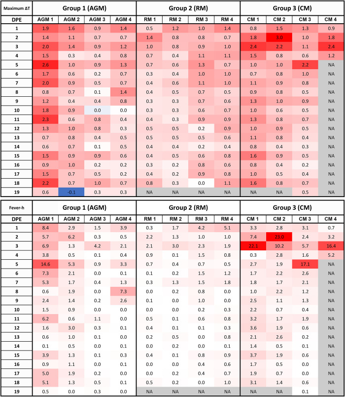
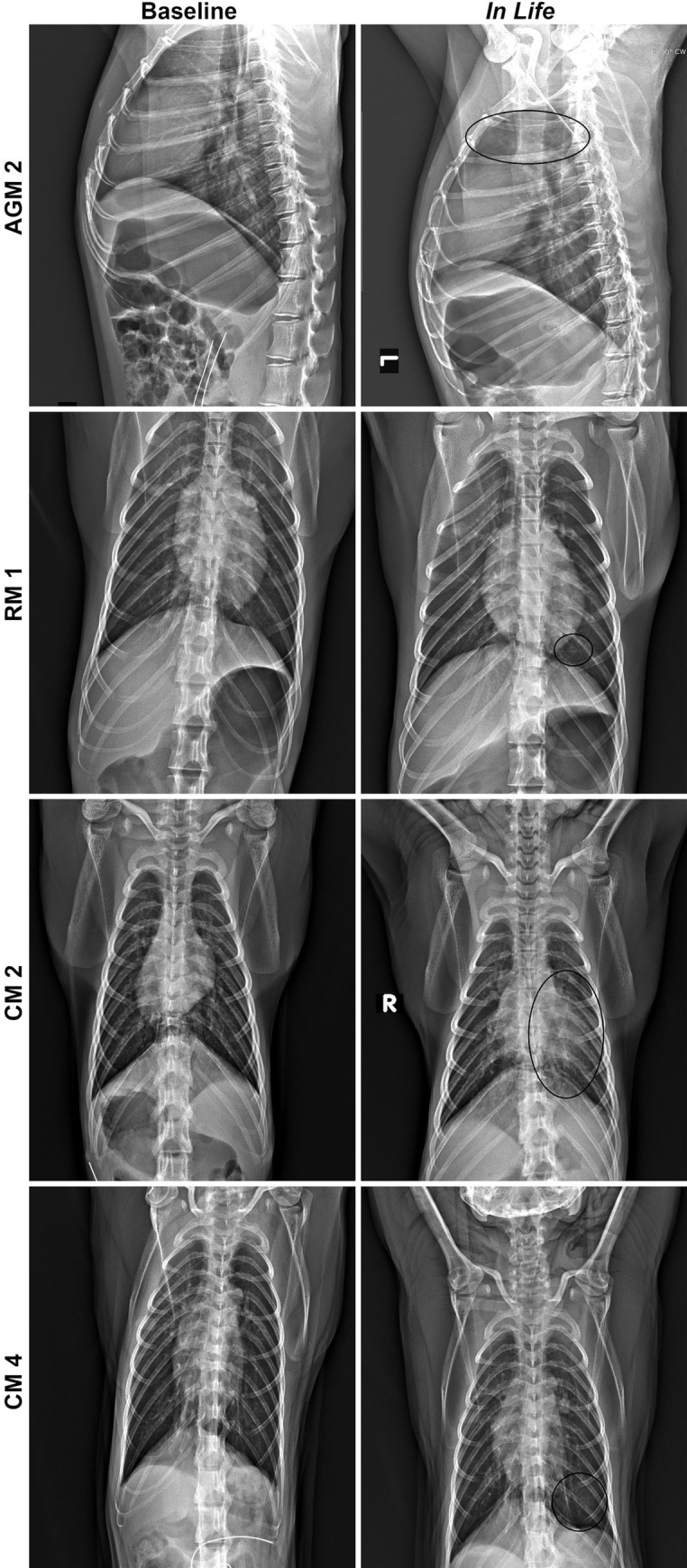
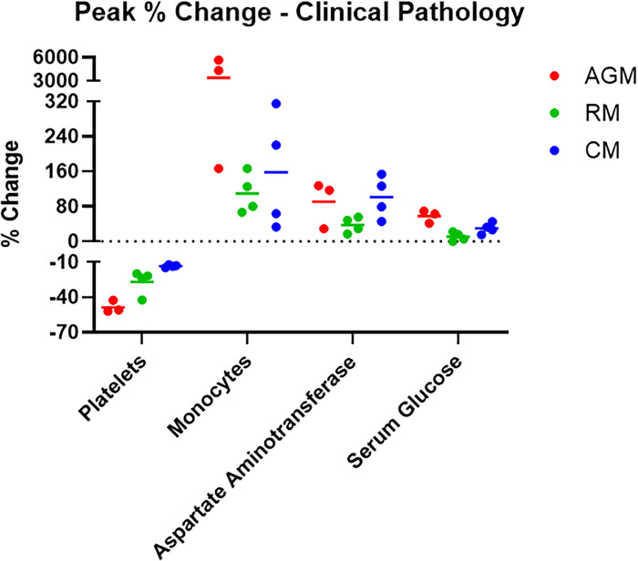
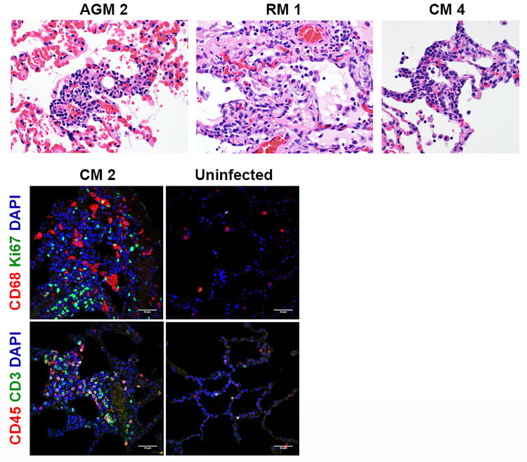
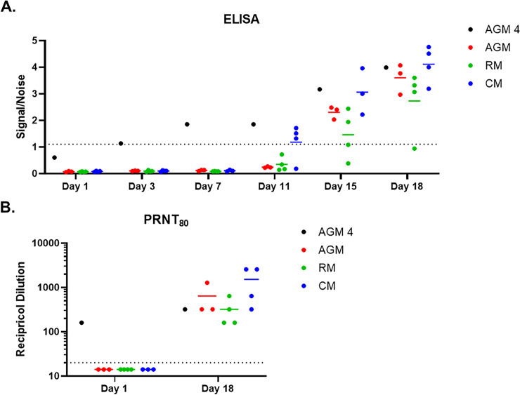
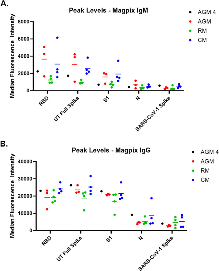
Airborne transmission is predicted to be a prevalent route of human exposure with SARS-CoV-2. Aside from African green monkeys, nonhuman primate models that replicate airborne transmission of SARS-CoV-2 have not been investigated. A comparative evaluation of COVID-19 in African green monkeys, rhesus macaques, and cynomolgus macaques following airborne exposure to SARS-CoV-2 was performed to determine critical disease parameters associated with disease progression, and establish correlations between primate and human COVID-19. Respiratory abnormalities and viral shedding were noted for all animals, indicating successful infection. Cynomolgus macaques developed fever, and thrombocytopenia was measured for African green monkeys and rhesus macaques. Type II pneumocyte hyperplasia and alveolar fibrosis were more frequently observed in lung tissue from cynomolgus macaques and African green monkeys. The data indicate that, in addition to African green monkeys, macaques can be successfully infected by airborne SARS-CoV-2, providing viable macaque natural transmission models for medical countermeasure evaluation.


Conflict of interest statement

The authors have declared that no competing interests exist.

Figures

Fig 1
Fig 1. Fever responses as measured by telemetry.
DSI M00 telemetry devices were used to collect body temperature data. The figure shows the maximum change in temperature (oC) for the 24 hour daily time period.
Fig 2
Fig 2. Radiographic changes for SARS-CoV-2 infected NHP.
Left panels show baseline images collected prior to telemetry surgery, and right panels show images collected on Study Day 7. All images are ventrodorsal except for AGM 2, which are lateral images. AGM 2 = Infiltrates present in cranial lung lobes (circled). RM 1 = Infiltrate present in caudal left lung (circled). CM 2 = Infiltrate present in left lung (circled). CM 4 = Increased opacity in caudal left lung (circled).
Fig 3
Fig 3. Hematology and clinical chemistries.
Hematology was performed on EDTA whole blood using a HM5 instrument. Clinical chemistries were performed on serum using a Piccolo Point-Of-Care instrument. Measurements for animals on study are shown as percent change from baseline (Study Day 1-challenge day values for that animal) for peak values for each analyte.
Fig 4
Fig 4. Viral RNA and live virus in oropharyngeal and nasopharyngeal swabs.
SARS-CoV-2-specific qRT-PCR (A-B) was performed on RNA extracted from nasopharyngeal and oropharyngeal swab clarified homogenates. Plaque assay (C-D) was performed on nasopharyngeal and oropharyngeal clarified homogenates. Data are shown as Log10 ge/mL (qRT-PCR) or Log10 pfu/mL (plaque assay), with nasopharyngeal swab data for each species shown in A and C, and oropharyngeal swab data for each species shown in B and D.
Fig 5
Fig 5. Histopathology and IFA.
Necropsies, histopathology, and IFA were performed on all animals. The top panels show the following histopathology findings: AGM 2 and CM 4 = type II pneumocyte hyperplasia; RM 1 = type II pneumocyte hyperplasia and alveolar fibrosis. The bottom panels show excessive CD68+ macrophages (red) and Ki67+ proliferating cells (green), CD45+ leukocytes (red), and CD3+ T cells infiltrated in alveolar septum for CM 2 compared to uninfected control lung tissue.
Fig 6
Fig 6. Characterization of immune response using ELISA and PRNT.
A. Serum samples were screened by commercial Euroimmun SARS-CoV-2 S1 IgG ELISA kit. This direct ELISA kit measures IgG response against the S1 subunit of the spike glycoprotein. The dotted line represents the assay cutoff, above which a sample is considered above background noise (i.e. positive). B. The PRNT80 titer is shown, and the dotted line represents the assay cutoff, above which a sample is considered above background noise (i.e. positive).
Fig 7
Fig 7. Characterization of immune response using a Magpix multiplex immunoassay.
Peak response for IgM (A) and IgG (B) to various antigens using the Magpix multiplex bead-based immunoassay is shown. Longitudinal Magpix data can be found in S3 Fig.
Fig 8
Fig 8. Viral RNA in nasopharyngeal swabs for AGM 4.
SARS-CoV-2-specific qRT-PCR was performed on RNA extracted from nasopharyngeal swab clarified homogenates. Data are shown as Log10 ge/mL. For comparison, data for AGM 1–3 are also shown.

References

    1. Chen N, Zhou M, Dong X, Qu J, Gong F, Han Y, et al. Epidemiological and clinical characteristics of 99 cases of 2019 novel coronavirus pneumonia in Wuhan, China: a descriptive study. Lancet. 2020;395(10223):507–13. Epub 2020/02/03. 10.1016/S0140-6736(20)30211-7 . - DOI - PMC - PubMed
    1. Huang C, Wang Y, Li X, Ren L, Zhao J, Hu Y, et al. Clinical features of patients infected with 2019 novel coronavirus in Wuhan, China. Lancet. 2020;395(10223):497–506. Epub 2020/01/28. 10.1016/S0140-6736(20)30183-5 . - DOI - PMC - PubMed
    1. Wang D, Hu B, Hu C, Zhu F, Liu X, Zhang J, et al. Clinical Characteristics of 138 Hospitalized Patients With 2019 Novel Coronavirus-Infected Pneumonia in Wuhan, China. JAMA. 2020. Epub 2020/02/08. 10.1001/jama.2020.1585 - DOI - PMC - PubMed
    1. Zhou F, Yu T, Du R, Fan G, Liu Y, Liu Z, et al. Clinical course and risk factors for mortality of adult inpatients with COVID-19 in Wuhan, China: a retrospective cohort study. Lancet. 2020. Epub 2020/03/15. 10.1016/S0140-6736(20)30566-3 . - DOI - PMC - PubMed
    1. Lu S, Zhao Y, Yu W, Yang Y, Gao J, Wang J, et al. Comparison of SARS-CoV-2 infections among 3 species of non-human primates. bioRxiv. 2020. Epub 2020/04/08. 10.1101/2020.04.08.031807 - DOI

Publication types