. 2021 Jan 26;12(2):164.
doi: 10.3390/genes12020164.

Evaluating the Role of Circulating Dendritic Cells in Methimazole-Treated Pediatric Graves' Disease Patients

Affiliations

Evaluating the Role of Circulating Dendritic Cells in Methimazole-Treated Pediatric Graves' Disease Patients

Aleksandra Starosz et al. Genes (Basel). .

Abstract

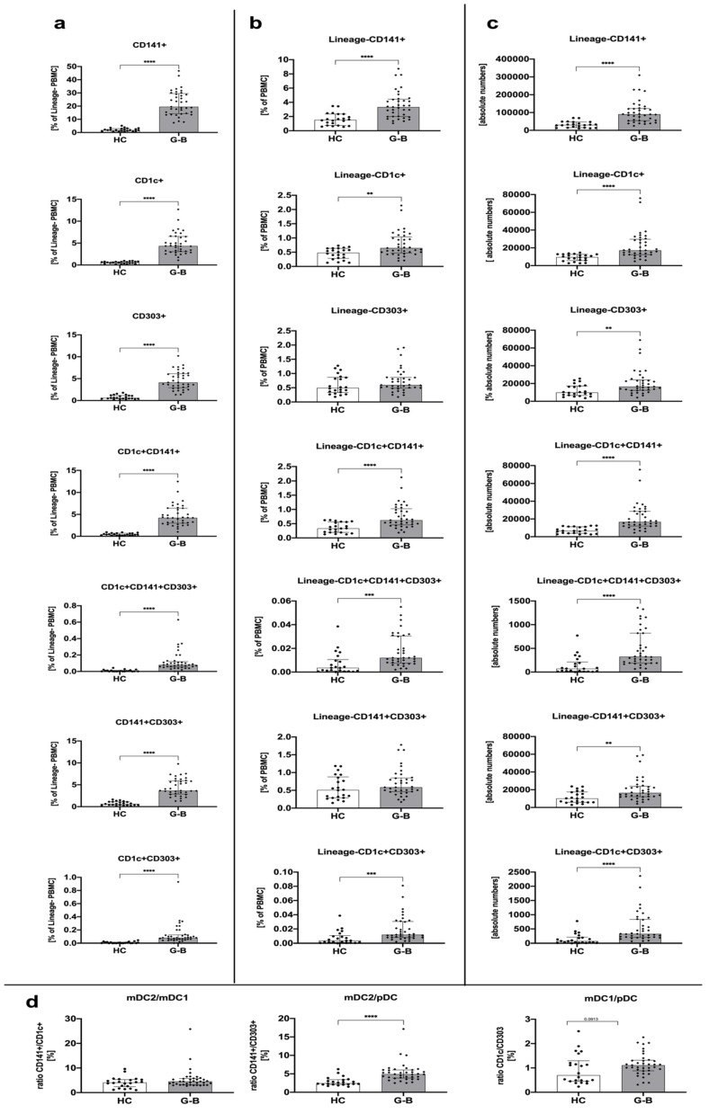
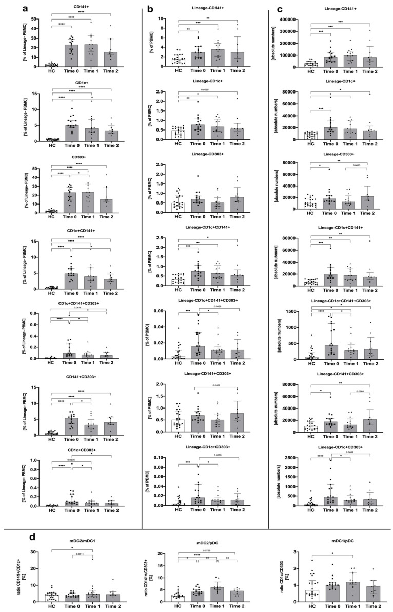
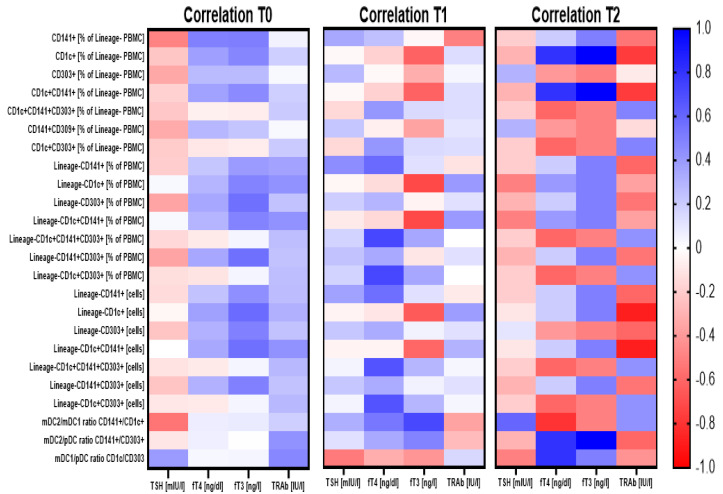
Graves' disease (GD) is hyperthyroidism associated with organ-specific autoimmune inflammation. GD occurs more frequently in adults than in children; however, pediatric patients are a therapeutic challenge due to cycles of remissions and relapses requiring constant monitoring at every stage of treatment administered. Dendritic cells (DCs) are considered to be a link between innate and adaptive immunity. DCs, as antigen-presenting cells (APCs), are involved in antigen presentation to T lymphocytes, thereby initiating a shift towards effector cells. In accordance, DCs also participate in the modulation of tolerance to specific antigens. To date, the data on DCs' role in Graves' pathological processes are scarce. Therefore, here, we evaluated the frequencies and role of circulating DCs in GD pediatric patients treated with methimazole. Flow cytometric analysis was implemented to evaluate three subsets of dendritic cells and their correlation with clinical GD-related parameters. We found significantly higher levels of DC subsets in patients at diagnosis. Furthermore, methimazole treatment seemed to effectively reduce subsets of DCs, which, in addition, were found to differentially correlate with thyroid function. Our study shed new light on DCs' role in the pediatric GD pathomechanism. Further studies are required for the mechanistic assessment of DCs' exact role in disease progression and influence on thyroid function.

Keywords: Graves’ disease; autoimmunity; dendritic cells; methimazole.


Conflict of interest statement

The authors declare no conflict of interest.

Figures

Figure 1
Figure 1
Dendritic cell-related differences among Graves’ disease patients (GD) and the healthy control group (HC). Acquired data were analyzed in the context of dendritic cell (DC) subset frequency within Lineage-negative peripheral blood mononuclear cells (PBMCs) (a), the total pool of PBMCs (b), as an absolute number of cells (c), and the ratio between studied DCs (d). Data are presented on each graph as the median with interquartile range. The levels of significant differences were indicated with: ** p < 0.01; *** p < 0.001; **** p < 0.0001.
Figure 2
Figure 2
Changes in dendritic cell-related parameters in the course of Graves’ disease patients’ treatment: before treatment (Time 0) and after 3 months (Time 1) and 1 year of treatment (Time 2). Acquired data were analyzed in the context of DC subset frequency within Lineage-negative PBMCs (a), the total pool of PBMCs (b), as an absolute number of cells (c), and the ratio between studied DCs (d). Data are presented on each graph as the median with interquartile range. The levels of significant differences were indicated with: * p < 0.05; ** p < 0.01; *** p < 0.001; **** p < 0.0001.
Figure 3
Figure 3
Demonstration of correlations between dendritic cell-related parameters and critical clinical analysis results prior to and during the treatment course. Correlation analysis demonstrated for three time points for Graves’ disease pediatric patients: before treatment (T0) and after 3 months (T1) and 1 year (T2) of the treatment. Strength of correlations (r value) are demonstrated through color gradient—blue for positive correlations and red for negative correlations.

Similar articles

Cited by

References

    1. Smith T.J., Hegedüs L. Graves’ Disease. N. Engl. J. Med. 2016;375:1552–1565. doi: 10.1056/NEJMra1510030. - DOI - PubMed
    1. Bossowski A.T., Reddy V., Perry L.A., Johnston L.B., Banerjee K., Blair J.C., Savage M.O. Clinical and endocrine features and long-term outcome of Graves’ disease in early childhood. J. Endocrinol. Investig. 2007;30:388–392. doi: 10.1007/BF03346315. - DOI - PubMed
    1. Kaguelidou F., Carel J.C., Léger J. Graves’ disease in childhood: Advances in management with antithyroid drug therapy. Horm. Res. 2009;71:310–317. doi: 10.1159/000223414. - DOI - PubMed
    1. Vita R., Lapa D., Trimarchi F., Vita G., Fallahi P., Antonelli A., Benvenga S. Certain HLA alleles are associated with stress-triggered Graves’ disease and influence its course. Endocrine. 2017;55:93–100. doi: 10.1007/s12020-016-0909-6. - DOI - PubMed
    1. Falgarone G., Heshmati H.M., Cohen R., Reach G. Mechanisms in endocrinology. Role of emotional stress in the pathophysiology of Graves’ disease. Eur. J. Endocrinol. 2013;168:R13–R18. doi: 10.1530/EJE-12-0539. - DOI - PubMed

Publication types