

Advanced Multi-Dimensional Cellular Models as Emerging Reality to Reproduce In Vitro the Human Body Complexity
- PMID: 33530487
- PMCID: PMC7865724
- DOI: 10.3390/ijms22031195
Advanced Multi-Dimensional Cellular Models as Emerging Reality to Reproduce In Vitro the Human Body Complexity
Abstract
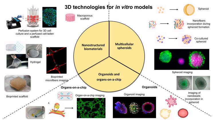
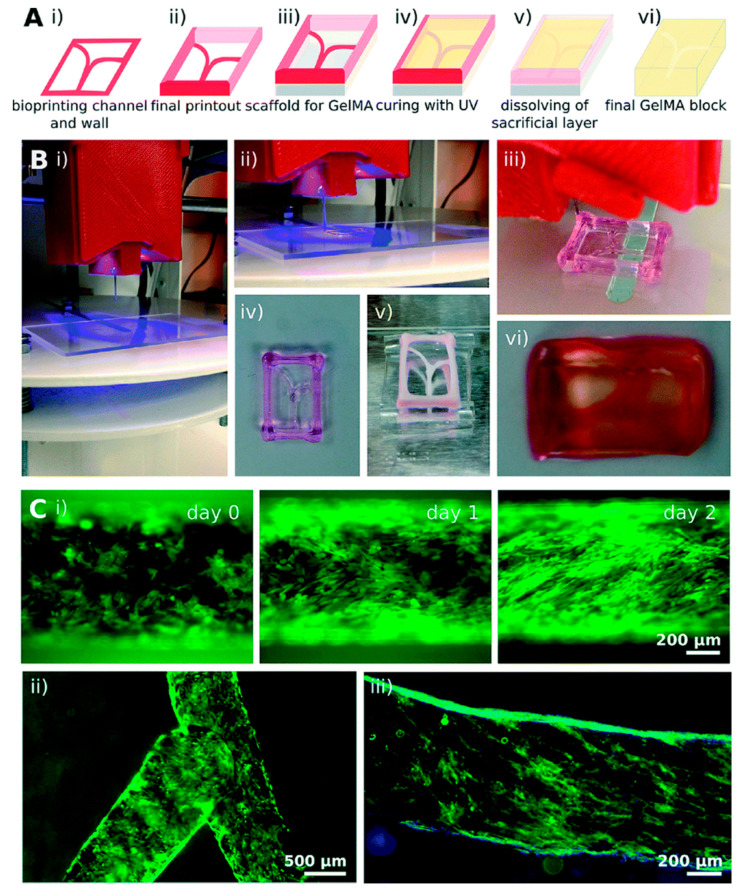
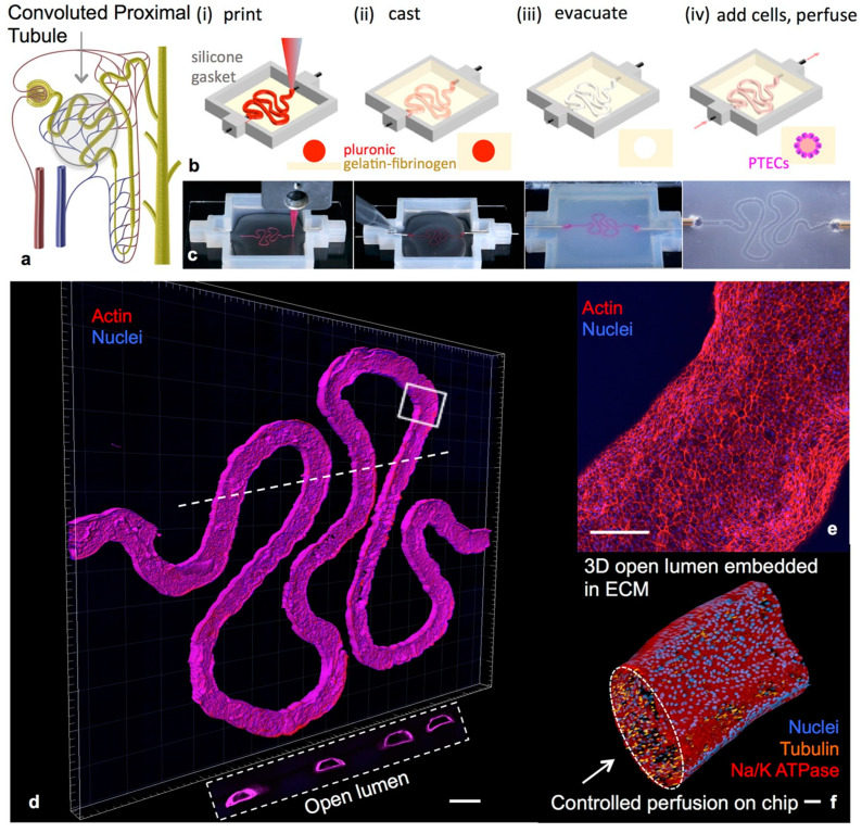
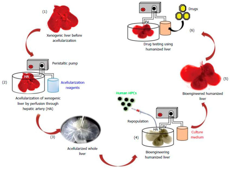
A hot topic in biomedical science is the implementation of more predictive in vitro models of human tissues to significantly improve the knowledge of physiological or pathological process, drugs discovery and screening. Bidimensional (2D) culture systems still represent good high-throughput options for basic research. Unfortunately, these systems are not able to recapitulate the in vivo three-dimensional (3D) environment of native tissues, resulting in a poor in vitro-in vivo translation. In addition, intra-species differences limited the use of animal data for predicting human responses, increasing in vivo preclinical failures and ethical concerns. Dealing with these challenges, in vitro 3D technological approaches were recently bioengineered as promising platforms able to closely capture the complexity of in vivo normal/pathological tissues. Potentially, such systems could resemble tissue-specific extracellular matrix (ECM), cell-cell and cell-ECM interactions and specific cell biological responses to mechanical and physical/chemical properties of the matrix. In this context, this review presents the state of the art of the most advanced progresses of the last years. A special attention to the emerging technologies for the development of human 3D disease-relevant and physiological models, varying from cell self-assembly (i.e., multicellular spheroids and organoids) to the use of biomaterials and microfluidic devices has been given.
Keywords: 3D in vitro models; multicellular spheroids; nanostructured biomaterials; organ-on-a-chip; organoids; tissue engineering.
Conflict of interest statement
The authors declare no conflict of interest.
Figures
References
-
- Ekert J.E., Deakyne J., Pribul-Allen P., Terry R., Schofield C., Jeong C.G., Storey J., Mohamet L., Francis J., Naidoo A., et al. Recommended Guidelines for Developing, Qualifying, and Implementing Complex In Vitro Models (CIVMs) for Drug Discovery. SLAS Discov. Adv. Life Sci. 2020 doi: 10.1177/2472555220923332. - DOI - PubMed
-
- Eurostat First Estimates of Research & Development Expenditure. R&D Expenditure in the EU Increased Slightly to 2.07% of GDP in 2017. [(accessed on 7 January 2019)]; Available online: https://ec.europa.eu/eurostat/documents/2995521/9483597/9-10012019-AP-EN....
-
- (PhRMA) Pharmaceutical Research and Manufacturers of America 2016 Biopharmaceutical Research Industry Profile. [(accessed on 8 January 2020)]; Available online: http://phrma-docs.phrma.org/sites/default/files/pdf/biopharmaceutical-in....
Publication types
MeSH terms
Substances
LinkOut - more resources
Full Text Sources
Other Literature Sources
