. 2021 Jan 26;22(3):1205.
doi: 10.3390/ijms22031205.

Hypoxia-Induced S100A8 Expression Activates Microglial Inflammation and Promotes Neuronal Apoptosis

Affiliations

Hypoxia-Induced S100A8 Expression Activates Microglial Inflammation and Promotes Neuronal Apoptosis

Ji Sun Ha et al. Int J Mol Sci. .

Abstract

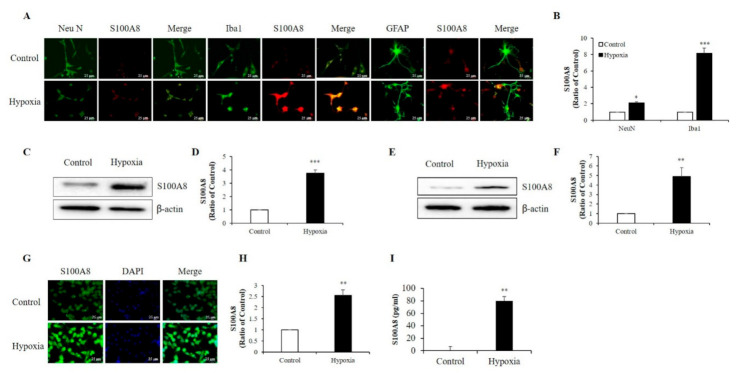
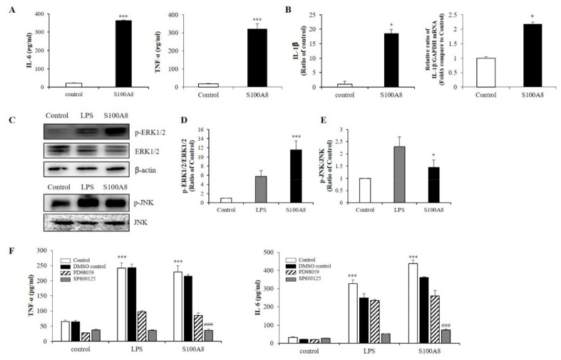
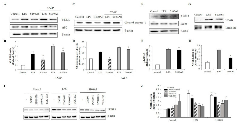
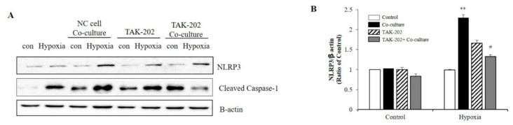
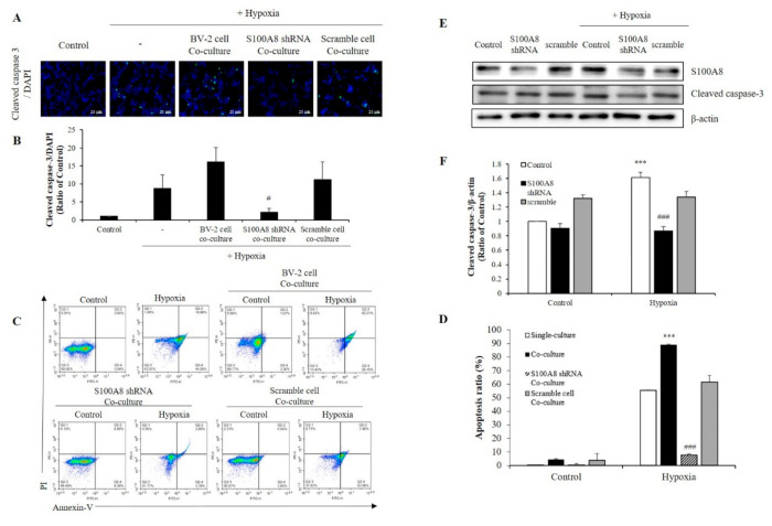
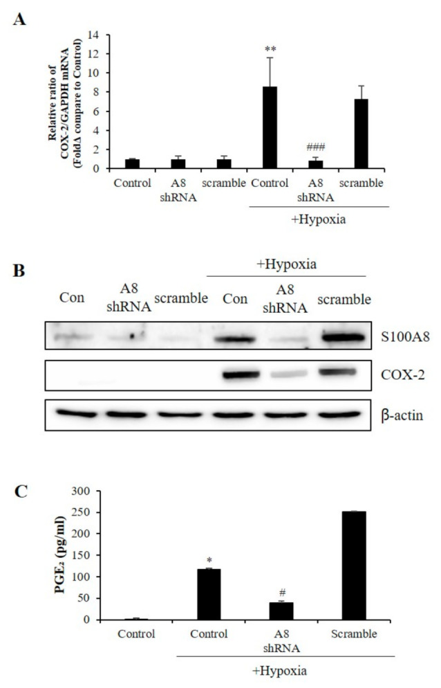
S100 calcium-binding protein A8 (S100A8), a danger-associated molecular pattern, has emerged as an important mediator of the pro-inflammatory response. Some S100 proteins play a prominent role in neuroinflammatory disorders and increase the secretion of pro-inflammatory cytokines in microglial cells. The aim of this study was to determine whether S100A8 induced neuronal apoptosis during cerebral hypoxia and elucidate its mechanism of action. In this study, we reported that the S100A8 protein expression was increased in mouse neuronal and microglial cells when exposed to hypoxia, and induced neuroinflammation and neuronal apoptosis. S100A8, secreted from neurons under hypoxia, activated the secretion of tumor necrosis factor (TNF-α) and interleukin-6 (IL-6) through phosphorylation of extracellular-signal-regulated kinase (ERK) and c-Jun N-terminal kinase (JNK) in microglia. Also, phosphorylation of ERK via the TLR4 receptor induced the priming of the NLRP3 inflammasome. The changes in Cyclooxygenase-2 (COX-2) expression, a well-known inflammatory activator, were regulated by the S100A8 expression in microglial cells. Knockdown of S100A8 levels by using shRNA revealed that microglial S100A8 expression activated COX-2 expression, leading to neuronal apoptosis under hypoxia. These results suggested that S100A8 may be an important molecule for bidirectional microglia-neuron communication and a new therapeutic target for neurological disorders caused by microglial inflammation during hypoxia.

Keywords: COX-2; S100A8; hypoxia; inflammasome; microglia; neuronal apoptosis.


Conflict of interest statement

The authors declare that they have no conflict of interest.

Figures

Figure 1
Figure 1
Hypoxia increased the production of S100 calcium-binding protein A8 (S100A8) in neuron and microglia and induced the release of S100A8 in SH-SY5Y cells. (A,B) S100A8 expression (red) were detected by immunocytochemical analysis in primary cultured neurons (NeuN, neuron marker) and cultured mixed glia (Iba1, microglial marker and GFAP, astrocyte marker) exposed to hypoxic conditions for 48 h. Scheme 25 μm. S100A8 expression was detected by western blot analysis in (C,D) SH-SY5Y cells and (E,F) BV-2 cells exposed to hypoxic conditions for 48 h. (G,H) S100A8 protein expression in BV-2 cells were confirmed by immunocytochemistry and (I) S100A8 release in SH-SY5Y was measured by enzyme-linked immunosorbent assay (ELISA) at 48 h after hypoxia. Values of * p < 0.05, ** p < 0.01, *** p < 0.001 versus control were considered as statistically significant.
Figure 2
Figure 2
S100A8 induces pro-inflammatory cytokines and inflammation in BV-2 cells. BV-2 cells were stimulated with S100A8 (10 μg/mL) for 24 h. (A) The supernatant was collected and TNF-α and interleukin-6 (IL-6) analyzed by ELISA. (B) The protein and mRNA were extracted, and the expression levels of IL-1β were assessed by ELISA and RT-qPCR. (CE) The protein was extracted, separated on 10% SDS-acrylamide gels (15 μg/lane) and transferred to nitrocellulose membrane. The protein expression level was detected by western blotting with anti-ERK1/2, anti-phospho-ERK1/2 (p-ERK1/2), anti-JNK and anti-p-JNK. (F) Cells were pre-treated with ERK inhibitor (PD98059, 20 μM), JNK inhibitor (SP600125, 10 μM) or the equivalent volume of DMSO for 1 h, then stimulated for 24 h with LPS or S100A8 for ELISA of TNF-α, IL-6. Data from three independent experiments are presented as the means ± S.D. Values of * p < 0.05, *** p < 0.001 versus control; ### p < 0.001 versus S100A8-treated sample were considered as statistically significant.
Figure 3
Figure 3
S100A8 induces inflammasome priming by toll-like receptor (TLR)-4 receptors associated with ERK and JNK pathway in BV-2 cells. BV-2 cells were incubated for 24 h with LPS (1 μg/mL) or S100A8 (10 μg/mL) followed by Adenosine 5′-triphosphate disodium salt hydrate (ATP) (1 mM) for 1 h. (A,B) The NLRP3, ASC, and (C,D) cleaved caspase-1 were detected by western blotting. β-actin was used as an internal control. (E,F) BV-2 cells were lysed to whole lysates and IκB-α phosphorylation was analyzed by western blotting. (G,H) The translocation of nuclear factor- κB (NF-κB) was also detected by western blotting. BV-2 cells were lysed to cytosolic extracts and nucleic extracts. Lamin-B1 was used as internal controls. (I,J) BV-2 microglial cells were pre-treated with PD98059 (ERK inhibitor, 20 μM), SP600125 (JNK inhibitor, 10 μM), TAK-202 (TLR4 inhibitor, 10 μg/mL) or an equivalent volume of DMSO and stimulated for 24 h with LPS or S100A8. Cells harvested and lysed in RIPA buffer for western blotting of NLRP3. Results are from one experiment that is representative of at least three others. Data from three independent experiments are presented as the means ± S.D. Values of * p < 0.05, ** p < 0.01 versus control; # p < 0.05, ## p < 0.01 versus S100A8-treated sample were considered as statistically significant.
Figure 4
Figure 4
S100A8 derived from neuronal cells induces NLRP3 inflammasome priming in microglia under hypoxic conditions. BV-2 cells were pre-treated with TAK-202 (TLR4 inhibitor, 10 μg/mL) for 1 h, then stimulated for 48 h in hypoxic condition with SH-SY5Y cells indirectly co-cultured in 0.4 μm pore transwell. (A) The protein expression level was detected by western blotting with NLRP3. β-actin was used as an internal control. (B) Quantitative analysis of NLRP3 levels. Data from three independent experiments are presented as the means ± S.D. Values of ** p < 0.01 versus control; # p < 0.05 versus co-cultured sample were considered as statistically significant.
Figure 5
Figure 5
The expression of S100A8 in microglial cell induces apoptosis of neuronal cells in hypoxic condition. (A,B) SH-SY5Y cells incubated without or with S100A8 KD BV-2 cells for 48 h in hypoxic condition. Cleaved caspase-3 immunofluorescence images and were detected and quantitative analysis of the number of cleaved-caspase3-positive cells are shown in lower panel. (C,D) Representative Annexin-V/PI images were detected by flow cytometry. Quantitative analysis of the apoptotic rate of SH-SY5Y cells are shown in lower panel. (E,F) Primary neuron-glial mixed cells were transfected with S100A8 shRNA vector for 24 h followed by 48 h in hypoxic condition. Cells were harvested, and the expression protein levels of S100A8 and cleaved caspase-3 were analyzed by Western blotting. Data from three independent experiments are presented as the means ± S.D. Values of *** p < 0.001 versus control; # p < 0.05, ### p < 0.001 versus hypoxia-exposed sample were considered as statistically significant.
Figure 6
Figure 6
The expression of S100A8 in microglial cell induces the Cyclooxygenase-2 (COX-2)/prostaglandin E2 (PGE2) pathway. BV-2 cells were transfected with S100A8 shRNA or Scramble vector. After 24 h, cells were incubated in hypoxic condition for 48 h. (A) The mRNA and (B) the protein levels of S100A8 and COX-2 were detected by real-time PCR and western blotting. (C) Secretion of PGE2 level analyzed by ELISA. Data from three independent experiments are presented as the means ± S.D. Values of * p < 0.05, ** p < 0.01 versus control; # p < 0.05, ### p < 0.001 versus hypoxia-exposed sample were considered as statistically significant.

References

    1. Johnson W., Onuma O., Owolabi M., Sachdev S. Stroke: A global response is needed. Bull. World Heal. Organ. 2016;94:634. doi: 10.2471/BLT.16.181636. - DOI - PMC - PubMed
    1. Lee Y., Lee S., Park J.-W., Hwang J.-S., Kim S.-M., Lyoo I.K., Lee C.-J., Han I.-O. Hypoxia-Induced Neuroinflammation and Learning–Memory Impairments in Adult Zebrafish Are Suppressed by Glucosamine. Mol. Neurobiol. 2018;55:8738–8753. doi: 10.1007/s12035-018-1017-9. - DOI - PubMed
    1. Khoshnam S.E., Winlow W., Farzaneh M., Farbood Y., Moghaddam H.F. Pathogenic mechanisms following ischemic stroke. Neurol. Sci. 2017;38:1167–1186. doi: 10.1007/s10072-017-2938-1. - DOI - PubMed
    1. Lo E.H., Dalkara T., Moskowitz M.A. Mechanisms, challenges and opportunities in stroke. Nat. Rev. Neurosci. 2003;4:399–414. doi: 10.1038/nrn1106. - DOI - PubMed
    1. Jayaraj R.L., Azimullah S., Beiram R., Jalal F.Y., Rosenberg G.A. Neuroinflammation: Friend and foe for ischemic stroke. J. Neuroinflamm. 2019;16:142. doi: 10.1186/s12974-019-1516-2. - DOI - PMC - PubMed

MeSH terms

LinkOut - more resources