. 2021 Jan 26;13(3):382.
doi: 10.3390/polym13030382.

Role of Curing Temperature of Poly(Glycerol Sebacate) Substrates on Protein-Cell Interaction and Early Cell Adhesion

Affiliations

Role of Curing Temperature of Poly(Glycerol Sebacate) Substrates on Protein-Cell Interaction and Early Cell Adhesion

Rubén Martín-Cabezuelo et al. Polymers (Basel). .

Abstract

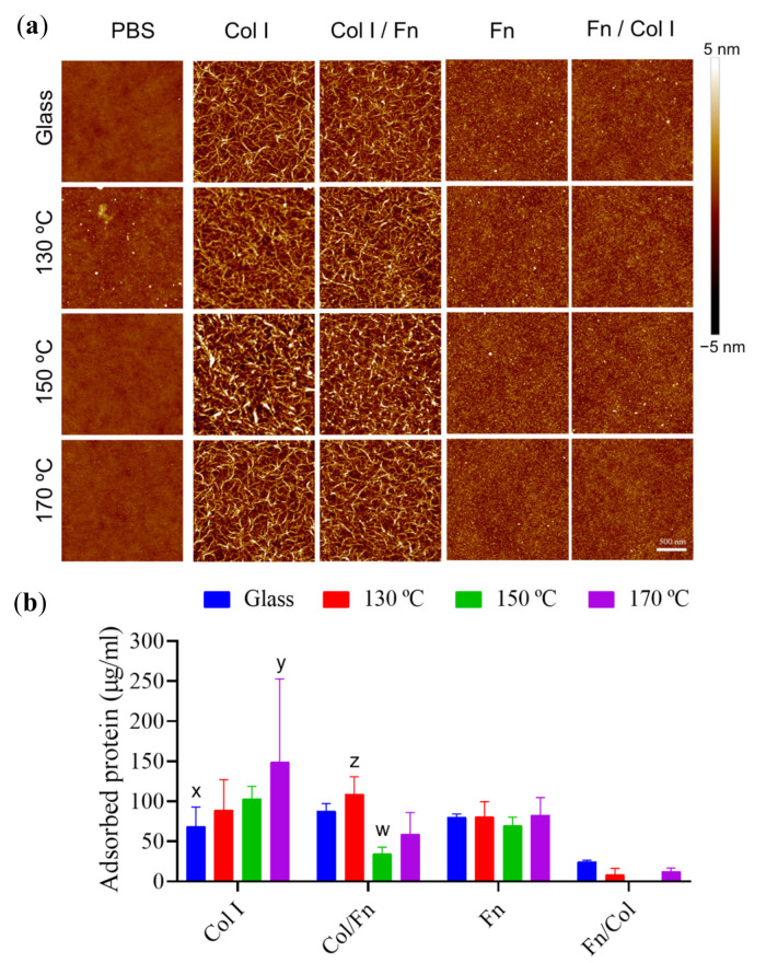
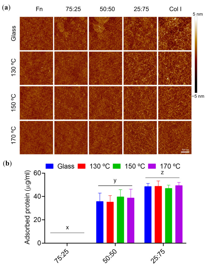
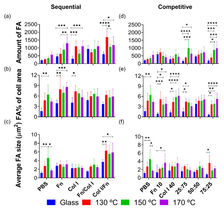
A novel procedure to obtain smooth, continuous polymeric surfaces from poly(glycerol sebacate) (PGS) has been developed with the spin-coating technique. This method proves useful for separating the effect of the chemistry and morphology of the networks (that can be obtained by varying the synthesis parameters) on cell-protein-substrate interactions from that of structural variables. Solutions of the PGS pre-polymer can be spin-coated, to then be cured. Curing under variable temperatures has been shown to lead to PGS networks with different chemical properties and topographies, conditioning their use as a biomaterial. Particularly, higher synthesis temperatures yield denser networks with fewer polar terminal groups available on the surface. Material-protein interactions were characterised by using extracellular matrix proteins such as fibronectin (Fn) and collagen type I (Col I), to unveil the biological interface profile of PGS substrates. To that end, atomic force microscopy (AFM) images and quantification of protein adsorbed in single, sequential and competitive protein incubations were used. Results reveal that Fn is adsorbed in the form of clusters, while Col I forms a characteristic fibrillar network. Fn has an inhibitory effect when incubated prior to Col I. Human umbilical endothelial cells (HUVECs) were also cultured on PGS surfaces to reveal the effect of synthesis temperature on cell behaviour. To this effect, early focal adhesions (FAs) were analysed using immunofluorescence techniques. In light of the results, 130 °C seems to be the optimal curing temperature since a preliminary treatment with Col I or a Fn:Col I solution facilitates the formation of early focal adhesions and growth of HUVECs.

Keywords: focal adhesion; poly(glycerol sebacate); polymer-protein interaction; protein adsorption.


Conflict of interest statement

The authors declare no conflict of interest. The funders had no role in the design of the study; nor in the collection, analyses, or interpretation of data; nor in the writing of the manuscript, nor in the decision to publish the results.

Figures

Figure 1
Figure 1
Chemical properties of poly(glycerol sebacate) (PGS) substrates. Fourier transform infrared spectroscopy (FTIR) spectra (transmittance) of PGS substrates obtained at different cured temperatures (130 °C, 150 °C and 170 °C) and glass cover slides. (a) Detailed FTIR spectra at the PGS characteristic chemical peak wavelengths: 1600–1800 cm-1 for COO peak on the right and 4000–2500 cm−1 for free –OH groups and -CH2 bonding on the left. (b) –OH/COO ratio obtained from transmittances at 3350 cm−1 and 2927 cm−1.
Figure 2
Figure 2
Human umbilical endothelial cells (HUVECs) biocompatibility on PGS substrates. HUVECs viability results from AlamarBlueTM colorimetry technique. Glass was used as a control of good cell viability, and a 10% DMSO solution was used as cytotoxic control. Sample data distributions were analysed through a two-way ANOVA and Bonferroni post hoc multiple mean comparison test with no significant differences between samples.
Figure 3
Figure 3
HUVECs FA quantification on PGS substrates. (a) Immunofluorescence images of HUVEC nuclei (cyan), actin cytoskeleton (green) and vinculin (magenta) as a focal adhesion (FA) marker of cells cultured after 3 h on glass and different PGS cured materials. Quantification of (b) the amount, (c) area and (d) average size of FA. FA area was calculated as the percentage of the total cell area. All immunofluorescence images share the same scale bar (20 µm). Sample data distributions were analysed through a one-way ANOVA and Bonferroni post hoc multiple mean comparison test, with a p-value < 0.05. *, p < 0.05 and ** p < 0.01.
Figure 4
Figure 4
Sequential protein adsorption on PGS substrates. (a) Atomic force microscope (AFM) height images of 2 × 2 µm2 and (b) protein adsorption quantification from microBCA colorimetric technique with single protein (Fn 100 μg/mL or Col I 400 μg/mL) and sequential adsorption assays of Fn and Col I (Fn 100 μg/mL+ Col I 80 μg/mL), and vice versa (Col I 400 μg/mL + Fn 20 μg/mL) on PGS cured at different temperatures. The first column in (a) shows the presence of PGS on the glass with no major difference in height for any of the applied curing temperatures. All images share the 500 nm scale bar. Glass covers without spin-coated PGS are shown in (b) as control. Sample data distributions were analysed through two-way ANOVA and a Bonferroni post hoc multiple mean comparison test, with a p-value of 0.01.
Figure 5
Figure 5
Protein competition adsorption on PGS substrates. (a) AFM height images of 2 × 2 µm2 and (b) protein adsorption quantification from microBCA colorimetric experiments after competition adsorption assays with Fn and Col I on glass and PGS cured at different temperatures. The indicated percentages of intermediate protein mixtures are volumetric ratios of Fn 10 µg/mL over Col I 40 µg/mL. All images share the scale bar of 500 nm. Sample data distributions were analysed through a two-way ANOVA and Bonferroni post hoc multiple mean comparison test, with a p-value of 0.0001.
Figure 6
Figure 6
FA parameters of HUVECs cultured on PGS substrates pre-treated in (ac) sequential and (df) competitive protein adsorption experiments. HUVECs fluorescence quantification of (a,d) number of FA, (b,e) relative surface of FA compared to the total cell area and (c,f) average size of FA, from protein sequential and competitive adsorption assays on glass and PGS cured at different temperatures. FA area was calculated as a percentage (%) of the total cell area. Sample data distributions were analysed through a two-way ANOVA and Bonferroni post hoc multiple mean comparison test, with a p-value < 0.05. *, p < 0.05; **, p < 0.01; ***, p < 0.001; and ****, p < 0.0001.

References

    1. Serrano M.C., Chung E.J., Ameer G.A. Advances and Applications of Biodegradable Elastomers in Regenerative Medicine. Adv. Funct. Mater. 2010;20:192–208. doi: 10.1002/adfm.200901040. - DOI
    1. Amsden B. Curable, biodegradable elastomers: Emerging biomaterials for drug delivery and tissue engineering. Soft Matter. 2007;3:1335. doi: 10.1039/b707472g. - DOI - PubMed
    1. Bat E., Zhang Z., Feijen J., Grijpma D.W., Poot A.A. Biodegradable elastomers for biomedical applications and regenerative medicine. Regen. Med. 2014;9:385–398. doi: 10.2217/rme.14.4. - DOI - PubMed
    1. Wang Y., Ameer G.A., Sheppard B.J., Langer R. A tough biodegradable elastomer. Nat. Biotechnol. 2002;20:602–606. doi: 10.1038/nbt0602-602. - DOI - PubMed
    1. Valerio O., Misra M., Mohanty A.K. Poly(glycerol- co-diacids) Polyesters: From Glycerol Biorefinery to Sustainable Engineering Applications, A Review. ACS Sustain. Chem. Eng. 2018;6:5681–5693. doi: 10.1021/acssuschemeng.7b04837. - DOI