. 2021 Jan 26;12(1):8.
doi: 10.3390/jfb12010008.

Poly(methyl methacrylate) Bone Cement Composite Can Be Refilled with Antibiotics after Implantation in Femur or Soft Tissue

Affiliations

Poly(methyl methacrylate) Bone Cement Composite Can Be Refilled with Antibiotics after Implantation in Femur or Soft Tissue

Erika L Cyphert et al. J Funct Biomater. .

Abstract

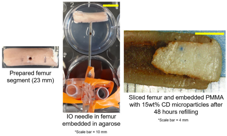
While periprosthetic joint infections (PJIs) result in a small percentage of patients following arthroplasties, they are challenging to treat if they spread into bone and soft tissue. Treatment involves delivering antibiotics using poly(methyl methacrylate) (PMMA) bone cement. However, antibiotic release is insufficient for prolonged infections. Previous work demonstrated efficacy of incorporating insoluble cyclodextrin (CD) microparticles into PMMA to improve antibiotic release and allow for post-implantation drug refilling to occur in a tissue-mimicking model. To simulate how antibiotic refilling may be possible in more physiologically relevant models, this work investigated development of bone and muscle refilling models. The bone refilling model involved embedding PMMA-CD into rabbit femur and administering antibiotic via intraosseous infusion. Muscle tissue refilling model involved implanting PMMA-CD beads in bovine muscle tissue and administering antibiotic via tissue injection. Duration of antimicrobial activity of refilled PMMA-CD was evaluated. PMMA-CD composite in bone and muscle tissue models was capable of being refilled with antibiotics and resulted in prolonged antimicrobial activity. PMMA-CD provided sustained and on-demand antimicrobial activity without removal of implant if infection develops. Intraosseous infusion appeared to be a viable technique to enable refilling of PMMA-CD after implantation in bone, reporting for the first time the ability to refill PMMA in bone.

Keywords: PMMA; antibiotic; bone cement; femur; refill.


Conflict of interest statement

The authors declare no conflict of interest.

Figures

Figure 1
Figure 1
Schematic of the setup of femur poly(methyl methacrylate) (PMMA)-cyclodextrin-(CD) composite antibiotic refilling model where PMMA containing CD microparticles was embedded in the bone and an intraosseous (IO) infusion needle was placed near the implanted PMMA-CD to serve as a port to inject antibiotics. Extent of antibiotic refilling of PMMA-CD using the model was evaluated through cutting the bone in half, explanting the PMMA-CD composite, imaging, and evaluating the duration of antimicrobial activity against S. aureus. Each experimental condition was evaluated in quadruplicate using this model.
Figure 2
Figure 2
Images of prepared femur segment with embedded PMMA-CD composite (left), femur segment embedded in agarose with IO needle inserted (middle), and interior of the femur segment 48 h following refilling with rifampicin (RMP) (right). Red/orange color along the periphery of the interior of the PMMA-CD was indicative of RMP refilling. Each experimental condition was evaluated in triplicate using this model.
Figure 3
Figure 3
Representative stereomicroscope images of the interior of PMMA-CD composite with and without (i.e., plain) 15 wt% CD microparticles after 48 h of refilling with RMP in femur model. Each experimental condition was evaluated in quadruplicate using this model with a minimum of eight measurements per sample.
Figure 4
Figure 4
Quantification of the depth or distance of diffusion of RMP into PMMA-CD composite with and without CD microparticles in the femur model and images depicting how representative measurements were collected. Red lines on photos indicate where diffusion was measured from. Each experimental condition was evaluated in quadruplicate using this model with a minimum of eight measurements per sample. ** Indicates p < 0.01. Ruler marks on images = 1 mm.
Figure 5
Figure 5
Persistence zone of inhibition study of PMMA-CD composite (with and without 15 wt% CD microparticles) refilled with RMP in femur model against S. aureus. Each experimental condition was evaluated in quadruplicate using this model with four measurements per sample. * Indicates p < 0.05.
Figure 6
Figure 6
Preparation of muscle tissue sample for implantation of PMMA-CD bead for antibiotic refilling and infection models. Success of antibiotic refilling of PMMA-CD bead through muscle tissue (with and without infection) was analyzed through persistence zone of inhibition studies and colony forming unit (CFU) counts. Each experimental condition was evaluated in triplicate using this model.
Figure 7
Figure 7
Images of implantation of PMMA-CD bead in muscle tissue in agarose, initial injection of RMP into muscle tissue, and muscle tissue following 48 h of incubation and refilling with RMP. Each experimental condition was evaluated in triplicate using this model.
Figure 8
Figure 8
Stereomicroscopy images of PMMA-CD beads after 1–3 cycles of RMP refilling in muscle tissue. Each experimental condition was evaluated in triplicate using this model.
Figure 9
Figure 9
Persistence zone of inhibition study of PMMA-CD beads after 1–3 cycles of RMP refilling in muscle tissue against S. aureus. Each experimental condition was evaluated in triplicate using this model with four measurements per sample.
Figure 10
Figure 10
Percent remaining CFU counts of S. aureus on PMMA-CD beads (with and without 15 wt% CD microparticles) and respective surrounding muscle tissue after treatment with RMP relative to no treatment. Each experimental condition was evaluated in triplicate using this model. * Indicates p < 0.05.
Figure 11
Figure 11
Persistence zone of inhibition study of PMMA-CD beads explanted from muscle tissue infection model after being refilled with RMP for 48 h. Each experimental condition was evaluated in triplicate using this model with four measurements per sample. * Indicates p < 0.05.

References

    1. Kaufman M., Meaike J., Izaddoost S. Orthopedic Prosthetic Infections: Diagnosis and Orthopedic Salvage. Semin. Plast. Surg. 2016;30:66–72. doi: 10.1055/s-0036-1580730. - DOI - PMC - PubMed
    1. McPherson E.J., Lewonowski K., Dorr L.D. Techniques in Arthroplasty: Use of an articulated PMMA spacer in the infected total knee arthroplasty. J. Arthroplast. 1995;10:87–89. doi: 10.1016/S0883-5403(05)80105-6. - DOI - PubMed
    1. Webb J.C.J., Spencer R.F. The role of polymethylmethacrylate bone cement in modern orthopaedic surgery. J. Bone Jt. Surg. 2007;89:851–857. doi: 10.1302/0301-620X.89B7.19148. - DOI - PubMed
    1. Bistolfi A., Massazza G., Verne E., Masse A., Deledda D., Ferraris S., Miola M., Galetto F., Crova M. Antibiotic-loaded cement in orthopedic surgery: A review. ISRN Orthop. 2011;2011:290851. doi: 10.5402/2011/290851. - DOI - PMC - PubMed
    1. Cyphert E.L., Learn G.D., Hurley S.K., Lu C.-Y., von Recum H.A. An additive to PMMA bone cement enables postimplantation drug refilling, broadens range of compatible antibiotics, and prolongs antimicrobial therapy. Adv. Healthc. Mater. 2018;7:1800812. doi: 10.1002/adhm.201800812. - DOI - PubMed

LinkOut - more resources