

Method for the Detection of the Cleaved Form of Shiga Toxin 2a Added to Normal Human Serum
- PMID: 33530614
- PMCID: PMC7911550
- DOI: 10.3390/toxins13020094
Method for the Detection of the Cleaved Form of Shiga Toxin 2a Added to Normal Human Serum
Abstract
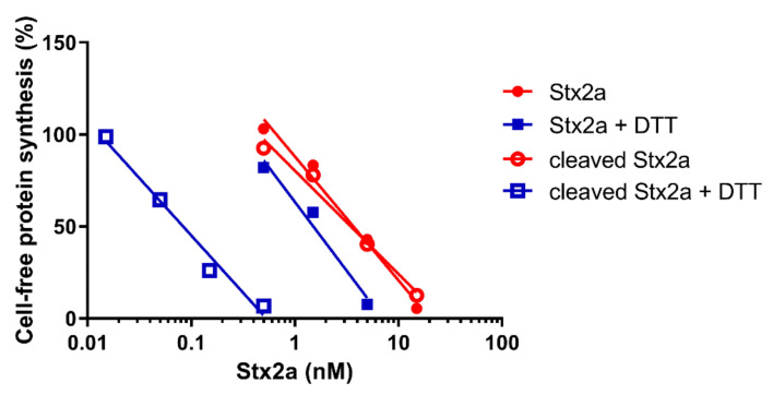
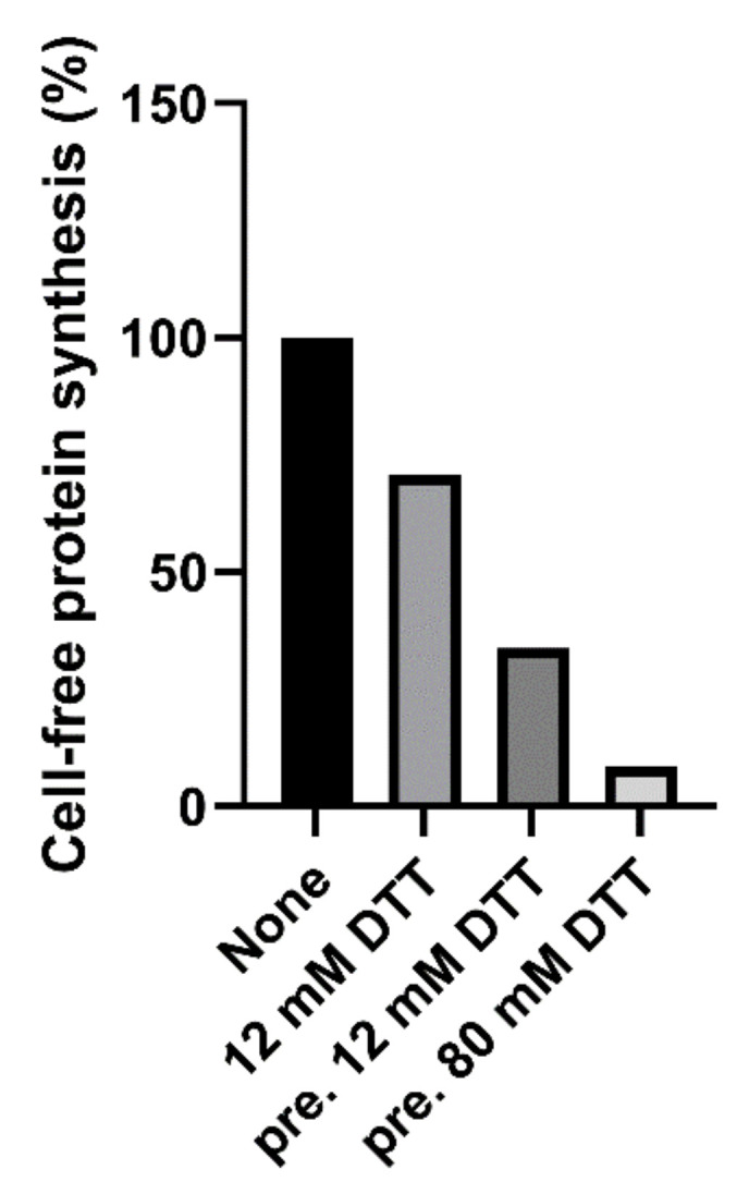
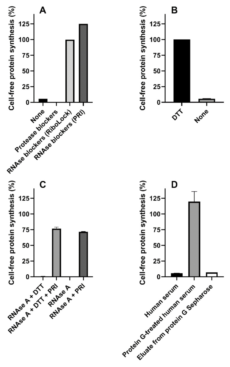
The pathogenesis of Escherichia coli-induced hemolytic uremic syndrome (eHUS) caused by infections with pathogenic Shiga toxin (Stx) producing E. coli (STEC) is centered on bacterial (e.g., Stx) and host factors (circulating cells, complement system, serum proteins) whose interaction is crucial for the immediate outcome and for the development of this life-threatening sequela. Stx2a, associated to circulating cells (early toxemia) or extracellular vesicles (late toxemia) in blood, is considered the main pathogenic factor in the development of eHUS. Recently, it was found that the functional properties of Stx2a (binding to circulating cells and complement components) change according to modifications of the structure of the toxin, i.e., after a single cleavage of the A subunit resulting in two fragments, A1 and A2, linked by a disulfide bridge. Herein, we describe a method to be used for the detection of the cleaved form of Stx2a in the serum of STEC-infected or eHUS patients. The method is based on the detection of the boosted inhibitory activity of the cleaved toxin, upon treatment with reducing agents, on a rabbit cell-free translation system reconstituted with human ribosomes. The method overcomes the technical problem caused by the presence of inhibitors of translation in human serum that have been stalled by the addition of RNAase blockers and by treatment with immobilized protein G. This method, allowing the detection of Stx2a at concentrations similar to those found by ELISA in the blood of STEC-infected patients, could be a useful tool to study the contribution of the cleaved form of Stx2a in the pathogenesis of eHUS.
Keywords: Shiga toxin-producing Escherichia coli; cleaved Shiga toxin 2a; hemolytic uremic syndrome.
Conflict of interest statement
The authors declare no conflict of interest.
Figures


References
-
- Brigotti M., He X., Carnicelli D., Arfilli V., Porcellini E., Galassi E., Tazzari P.L., Ricci F., Patfield S.A., Testa S., et al. Particulate Shiga toxin 2 in blood is associated to the development of hemolytic uremic syndrome in children. Thromb. Haemost. 2020;120:107–120. doi: 10.1055/s-0039-3400301. - DOI - PubMed
-
- Stahl A.L., Arvidsson I., Johansson K.E., Chromek M., Rebetz J., Loos S., Kristoffersson A.C., Bekassy Z.D., Morgelin M., Karpman D. A novel mechanism of bacterial toxin transfer within host blood cell-derived microvesicles. PLoS Pathog. 2015;11:e1004619. doi: 10.1371/journal.ppat.1004619. - DOI - PMC - PubMed
Publication types
MeSH terms
Substances
Grants and funding
LinkOut - more resources
Full Text Sources
Other Literature Sources
Medical
