. 2021 Jan 26;12(2):170.
doi: 10.3390/genes12020170.

The Axenfeld-Rieger Syndrome Gene FOXC1 Contributes to Left-Right Patterning

Affiliations

The Axenfeld-Rieger Syndrome Gene FOXC1 Contributes to Left-Right Patterning

Paul W Chrystal et al. Genes (Basel). .

Abstract

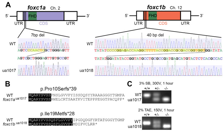
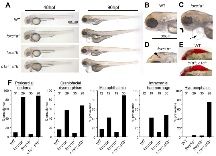
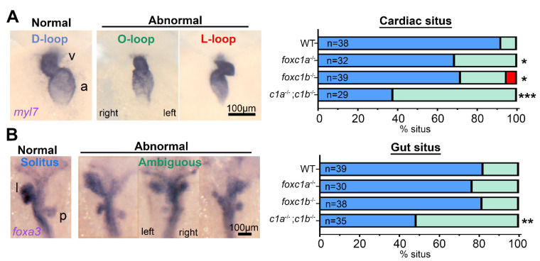
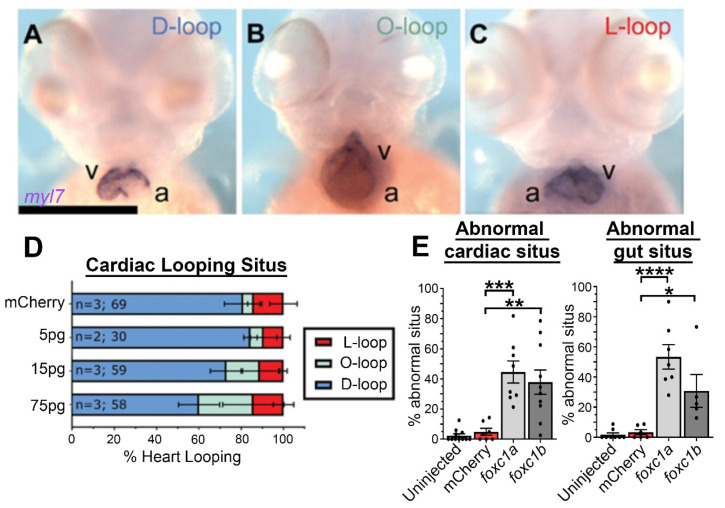
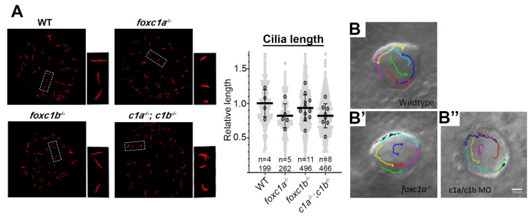
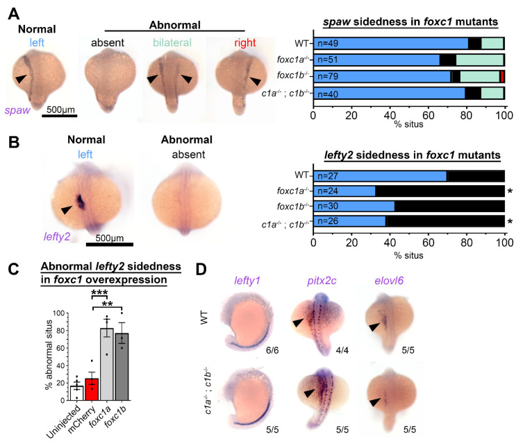
Precise spatiotemporal expression of the Nodal-Lefty-Pitx2 cascade in the lateral plate mesoderm establishes the left-right axis, which provides vital cues for correct organ formation and function. Mutations of one cascade constituent PITX2 and, separately, the Forkhead transcription factor FOXC1 independently cause a multi-system disorder known as Axenfeld-Rieger syndrome (ARS). Since cardiac involvement is an established ARS phenotype and because disrupted left-right patterning can cause congenital heart defects, we investigated in zebrafish whether foxc1 contributes to organ laterality or situs. We demonstrate that CRISPR/Cas9-generated foxc1a and foxc1b mutants exhibit abnormal cardiac looping and that the prevalence of cardiac situs defects is increased in foxc1a-/-; foxc1b-/- homozygotes. Similarly, double homozygotes exhibit isomerism of the liver and pancreas, which are key features of abnormal gut situs. Placement of the asymmetric visceral organs relative to the midline was also perturbed by mRNA overexpression of foxc1a and foxc1b. In addition, an analysis of the left-right patterning components, identified in the lateral plate mesoderm of foxc1 mutants, reduced or abolished the expression of the NODAL antagonist lefty2. Together, these data reveal a novel contribution from foxc1 to left-right patterning, demonstrating that this role is sensitive to foxc1 gene dosage, and provide a plausible mechanism for the incidence of congenital heart defects in Axenfeld-Rieger syndrome patients.

Keywords: Axenfeld–Rieger syndrome; FOXC1; LEFTY; left–right patterning; zebrafish.


Conflict of interest statement

The authors declare no conflict of interest.

Figures

Figure 1
Figure 1
Allelic consequence of foxc1-targeted CRISPR/Cas9 mutagenesis: (A) a schematic representation of foxc1aua1017 and foxc1bua1018 mutations, with 7 and 40 base pair deletions (yellow highlighting) upstream of the DNA-binding Forkhead domain; (B) the predicted sequences of the first 40 amino acids translated from wildtype (WT) and mutant proteins with sequence identity (black highlighting), where the foxc1aua1017 allele produces a truncated 39-residue protein with loss of sequence homology from amino acid 10 and the foxc1bua1018 allele produces a truncated 28-residue protein with loss of sequence homology from amino acid 19; and (C) PCR genotyping from the gDNA template resolving the respective deletions in foxc1a and foxc1b. (FHD, Forkhead domain; UTR, untranslated region; CDS, coding sequence.).
Figure 2
Figure 2
Foxc1 mutants exhibit multiple developmental defects. (A) foxc1 single mutants are largely indistinguishable from WT (wildtype) controls at 48 hpf (left panels), whereas foxc1a−/−; foxc1b−/− double homozygotes display hydrocephalus and oedema. By 96 hpf, no changes are observed in foxc1b−/− homozygotes, however; foxc1a−/− homozygotes and foxc1a−/−; foxc1b−/− double homozygotes display pericardial oedema (compare B and C, as highlighted by the arrow), microphthalmia (dotted circle), and craniofacial dysmorphism (chevron). At this stage, a subset of mutants also displayed intracranial hemorrhage (arrowhead in panel D), with foxc1a−/− homozygotes having greater frequency than foxc1b−/− homozygotes (42% vs. 11%, respectively), and (E) foxc1a−/−; foxc1b−/− double homozygotes present with hydrocephalus. (F) Quantification reveals that these defects are incompletely penetrant and generally more prevalent in double than single homozygotes (the number of embryos analyzed is shown above each bar). In the case of hydrocephalus, only the double homozygotes display an appreciable frequency of this phenotype.
Figure 3
Figure 3
Decreased dosages of foxc1 result in multi-organ situs defects. (A) In situ hybridization with myl7 at 48 hpf revealed aberrant cardiac situs (O-loop (green); L-loop (red)) compared with normal D-loop (blue). The prevalence of aberrant situs is increased in foxc1a, foxc1b, and double homozygotes (* p = 0.016, * 0.036, *** < 0.001 respectively, Fisher’s exact test) when compared to WT siblings. (B) At the same stage, the normal arrangement (solitus, blue) of the gut is left-sided liver (l) and right-sided pancreas (p); however, the incidence of abnormal (ambiguous, green) gut situs was significantly greater in double homozygotes (** p = 0.003, Fisher’s exact test).
Figure 4
Figure 4
Foxc1a mRNA overexpression causes cardiac situs defects in a dose-dependent manner: (AC) myl7 in situ hybridization of 75 pg foxc1a mRNA-injected embryos at 48 hpf, with representative images of the three cardiac looping morphologies provided (v, ventricle; a, atrium); (D) quantification of cardiac situs in control and foxc1a mRNA-injected embryos revealing an increasing prevalence of anomalous cardiac looping with increasing amounts of foxc1a mRNA; and (E) quantification of embryo situs defects in embryos injected with 75 pg of foxc1a mRNA. Statistical significance was apparent in comparisons between foxc1a/b and mCherry controls (cardiac: p = 0.0002 and 0.0012; gut: p < 0.0001 and 0.0205. mCherry vs. foxc1a and foxc1b respectively. One-way ANOVA and Dunnett’s post hoc test. * p < 0.05, ** p < 0.01, *** p < 0.001, **** p < 0.0001).
Figure 5
Figure 5
Loss of foxc1 does not significantly change cilia length or left–right organizer flow. (A) Acetylated α-tubulin immunostaining of left–right organizer (LRO) cilia revealed that average cilia length was not significantly altered in foxc1 mutants (foxc1a−/−, 82%; foxc1b−/−, 93%; foxc1a−/−; foxc1b−/−, 82% of WT length; p = 0.286, ANOVA; 4–11 embryos imaged per condition, all cilia per condition shown in light grey). Tracking of fluorescent bead flow in the LRO revealed unchanged counterclockwise flow between WT (B), foxc1a−/− homozygotes (B’), and foxc1a/foxc1b morphants (B’’).
Figure 6
Figure 6
Foxc1 mutants display loss of lefty2 expression independent of changes to other left–right patterning genes. (A) In situ hybridization with southpaw (spaw) revealed no difference in the prevalence of normal left-sided expression (normal = blue, absent = black, bilateral = green, and right = red) in foxc1 mutants compared to controls (p > 0.1, Fisher’s exact test). (B) Conversely, lefty2 expression was significantly altered in foxc1 mutants. Normal left-sided lefty2 expression was absent more frequently in foxc1a−/− and double foxc1a−/−; foxc1b−/− homozygotes (p = 0.012 and 0.028, respectively) and trended to be absent in foxc1b−/− homozygotes without reaching statistical significance (p = 0.061, Fisher’s exact test) (normal = blue and absent = black). (C) lefty2 expression was significantly abnormal, with the overexpression of foxc1a or foxc1b (p = 0.0007 and 0.0030, respectively, ANOVA and Dunnett’s Test). (D) Analysis of the additional left–right patterning genes lefty1, pitx2c, and elvol6 revealed no differences between WT, and foxc1a−/−; foxc1b−/− double homozygotes. (* p < 0.05, ** p < 0.01, *** p < 0.001).

References

    1. Peeters H., Devriendt K. Human laterality disorders. Eur. J. Med. Genet. 2006;49:349–362. doi: 10.1016/j.ejmg.2005.12.003. - DOI - PubMed
    1. Sutherland M.J., Ware S.M. Disorders of left-right asymmetry: Heterotaxy and situs inversus. Am. J. Med. Genet. C Semin. Med. Genet. 2009;151C:307–317. doi: 10.1002/ajmg.c.30228. - DOI - PubMed
    1. Shiraishi I., Ichikawa H. Human heterotaxy syndrome—From molecular genetics to clinical features, management, and prognosis. Circ. J. 2012;76:2066–2075. doi: 10.1253/circj.CJ-12-0957. - DOI - PubMed
    1. Taketazu M., Lougheed J., Yoo S.J., Lim J.S., Hornberger L.K. Spectrum of cardiovascular disease, accuracy of diagnosis, and outcome in fetal heterotaxy syndrome. Am. J. Cardiol. 2006;97:720–724. doi: 10.1016/j.amjcard.2005.09.119. - DOI - PubMed
    1. Borenstein S.H., Langer J.C. Heterotaxia syndromes and their abdominal manifestations. Curr. Opin. Pediatr. 2006;18:294–297. doi: 10.1097/01.mop.0000193313.75827.e9. - DOI - PubMed

Publication types

MeSH terms

Substances

Supplementary concepts

LinkOut - more resources