

Trans-Tubular Translaminar Microscopic-Assisted Nucleotomy for Lumbar Disc Herniations in the Hidden Zone
- PMID: 33530710
- PMCID: PMC9393970
- DOI: 10.1177/2192568221990421
Trans-Tubular Translaminar Microscopic-Assisted Nucleotomy for Lumbar Disc Herniations in the Hidden Zone
Erratum in
-
Corrigendum to "Trans-Tubular Translaminar Microscopic-Assisted Nucleotomy for Lumbar Disc Herniations in the Hidden Zone".Global Spine J. 2022 Jul;12(6):1311. doi: 10.1177/21925682221078322. Epub 2022 Mar 29. Global Spine J. 2022. PMID: 35350903 Free PMC article. No abstract available.
Abstract
Study design: A prospective cohort study in a high-flow spine center in Germany.
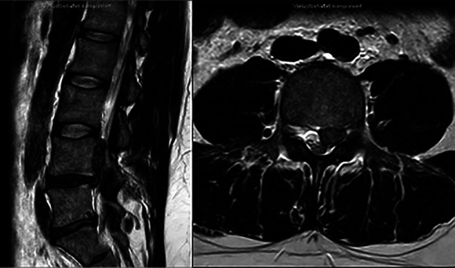
Objectives: This study aimed to evaluate clinical outcomes and complications of the trans-tubular translaminar microscopic-assisted percutaneous nucleotomy in cases of cranially migrated lumbar disc herniations (LDH).
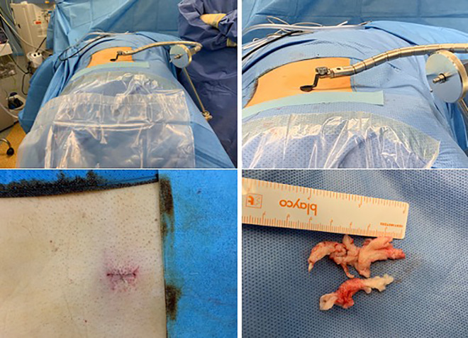
Methods: Between January 2013 and January 2018, 66 consecutive patients with cranio-laterally migrated LDH were operated upon. The following outcome measures were evaluated: (1) Visual Analog Scale (VAS) for leg and back pain; (2) Oswestry Disability Index (ODI) and Macnab´s criteria. All patients were operated upon with trans-tubular Translaminar Microscopic-assisted Percutaneous Nucleotomy (TL-MAPN). Perioperative radiographic and clinical evaluations were reported. The mean follow-up period was 32 months.
Results: The mean age was 59 years. L4/L5 was the commonest affected level (27 patients). The mean preoperative VAS for leg pain was 6.44 (±2.06), improved to 0,35 (±0.59) postoperatively. Dural injury occurred in 1 patient, treated with dural patch. Improved neurological function was reported in 41/44 Patients (neurological improvement rate of 93%) at the final follow up. There was a significant improvement in the mean ODI values, from 50.19 ± 4.92 preoperatively to 10.14 ± 2.22 postoperatively (P < 0.001). Sixty four out of 66 patients (96%) showed an excellent or good functional outcome according to Macnab´s criteria. No recurrent herniations were observed.
Conclusion: The translaminar approach is a viable minimal invasive technique for cranially migrated LDH. The preservation of the flavum ligament is one of the main advantages of this technique. It is an effective, safe and reproducible minimally invasive surgical alternative in treatment of cranially migrated LDHs.
Keywords: cranially migrated disc; discectomy; lumbar disc herniation; minimal invasive; trans-laminar trans-tubular.
Conflict of interest statement
Figures


Similar articles
-
Extraforaminal microscopic assisted percutaneous nucleotomy for foraminal and extraforaminal lumbar disc herniations.Spine J. 2018 Apr;18(4):620-625. doi: 10.1016/j.spinee.2017.08.258. Epub 2017 Sep 4. Spine J. 2018. PMID: 28882526
-
Radiographic and clinical outcomes of huge lumbar disc herniations treated by transforaminal endoscopic discectomy.Clin Neurol Neurosurg. 2019 Oct;185:105485. doi: 10.1016/j.clineuro.2019.105485. Epub 2019 Aug 9. Clin Neurol Neurosurg. 2019. PMID: 31421587
-
The Use of Tubular Retractors for Translaminar Discectomy for Cranially and Caudally Extruded Discs.Indian J Orthop. 2018 May-Jun;52(3):328-333. doi: 10.4103/ortho.IJOrtho_364_16. Indian J Orthop. 2018. PMID: 29887637 Free PMC article.
-
Percutaneous endoscopic interlaminar discectomy via inner border of inferior pedicle approach for downmigrated disc herniation: a retrospective study.J Orthop Surg Res. 2022 Jul 21;17(1):359. doi: 10.1186/s13018-022-03245-8. J Orthop Surg Res. 2022. PMID: 35864515 Free PMC article. Review.
-
Technical note: microdiscectomy and translaminar approach.J Spine Surg. 2015 Dec;1(1):44-9. doi: 10.3978/j.issn.2414-469X.2015.10.03. J Spine Surg. 2015. PMID: 27683678 Free PMC article. Review.
Cited by
-
Complications in Minimally Invasive Spine Surgery in the Last 10 Years: A Narrative Review.Neurospine. 2024 Sep;21(3):770-803. doi: 10.14245/ns.2448652.326. Epub 2024 Sep 30. Neurospine. 2024. PMID: 39363458 Free PMC article.
References
-
- Soldner F, Hoelper B, Wallenfang T, Behr R. The translaminar approach to canalicular and cranio-dorsolateral lumbar disc herniations. Acta Neurochir. 2002;144(4):315–320. - PubMed
-
- Bernucci C, Giovanelli M. Translaminar microsurgical approach for lumbar herniated nucleus pulposus (HNP) in the “hidden zone”: clinical and radiologic results in a series of 24 patients. Spine. 2007;32(2):281–284. - PubMed
-
- Papavero L, Langer N, Fritzsche E, Emami P, Westphal M, Kothe R. The translaminar approach to lumbar disc herniations impinging the exiting root. Operative Neurosurgery. 2008;62(suppl_1): ONS173–ONS178. - PubMed
-
- Macnab I. Negative disc exploration: an analysis of the causes of nerve-root involvement in sixty-eight patients. J Bone Joint Surg Am. 1971;53(5):891–903. - PubMed
-
- Faulhauer K, Manicke C. Fragment excision versus conventional disc removal in the microsurgical treatment of herniated lumbar disc. Acta Neurochir (Wien). 1995;133(3-4):107–111. - PubMed
LinkOut - more resources
Full Text Sources
Other Literature Sources
