

Successful colonoscopic removal of a foreign body that caused sigmoid colon perforation: a case report
- PMID: 33530808
- PMCID: PMC7871075
- DOI: 10.1177/0300060520982828
Successful colonoscopic removal of a foreign body that caused sigmoid colon perforation: a case report
Abstract
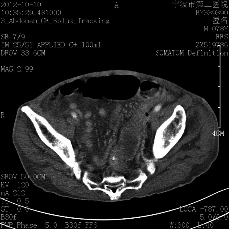
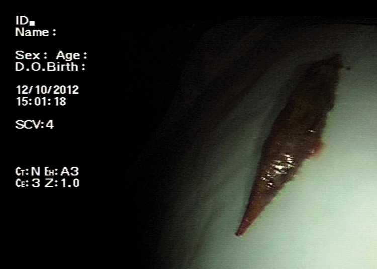
Large bowel perforation is an acute abdominal emergency requiring rapid diagnosis for proper treatment. The high mortality rate associated with large bowel perforation underlines the importance of an accurate and timely diagnosis. Computed tomography is useful for diagnosis of ingested foreign bodies, and endoscopic repair using clips can be an effective treatment of colon perforations. We herein describe a 78-year-old man with sigmoid colon perforation caused by accidental swallowing of a jujube pit. The jujube pit had become stuck in the wall of the sigmoid colon and was successfully removed by colonoscopy, avoiding an aggressive surgery. As a result of developments in endoscopic techniques, endoscopic closure has become a feasible option for the management of intestinal perforation.
Keywords: Perforation; colonoscopy; computed tomography; endoscopic repair; foreign body; jujube.
Conflict of interest statement
Figures


References
-
- Bielecki K, Kaminski P, Klukowski M. Large bowel perforation: morbidity and mortality. Tech Coloproctol 2002; 6: 177–182. - PubMed
-
- Kriwanek S, Armbruster C, Beckerhinn P, et al. Prognostic factors for survival in colonic perforation. Int J Colorectal Dis 1994; 9: 158–162. - PubMed
-
- Kim JS, Kim BW, Kim JI, et al. Endoscopic clip closure versus surgery for the treatment of iatrogenic colon perforations developed during diagnostic colonoscopy: a review of 115,285 patients. Surg Endosc 2013; 27: 501–504. - PubMed
-
- Pinero Madrona A, Fernandez Hernandez JA, Carrasco Prats M, et al . Intestinal perforation by foreign bodies. Eur J Surg 2000; 166: 307–309. - PubMed
Publication types
MeSH terms
LinkOut - more resources
Full Text Sources
Other Literature Sources
Medical
Research Materials
