Comparative Study
. 2021 Feb 2;20(1):33.
doi: 10.1186/s12934-021-01528-z.

Decorating the surface of Escherichia coli with bacterial lipoproteins: a comparative analysis of different display systems

Affiliations
Comparative Study

Decorating the surface of Escherichia coli with bacterial lipoproteins: a comparative analysis of different display systems

Sonia Nicchi et al. Microb Cell Fact. .

Abstract

Background: The display of recombinant proteins on cell surfaces has a plethora of applications including vaccine development, screening of peptide libraries, whole-cell biocatalysts and biosensor development for diagnostic, industrial or environmental purposes. In the last decades, a wide variety of surface display systems have been developed for the exposure of recombinant proteins on the surface of Escherichia coli, such as autotransporters and outer membrane proteins.

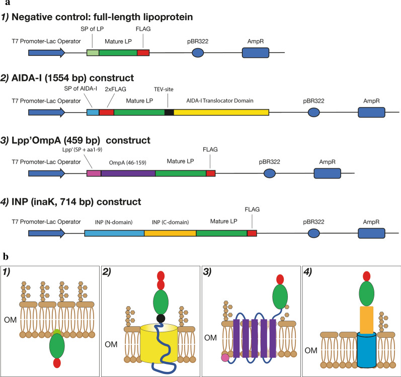
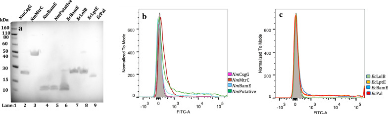
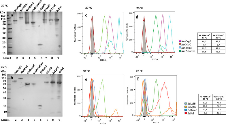
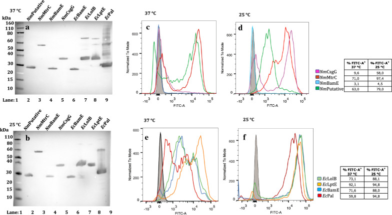
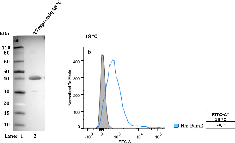
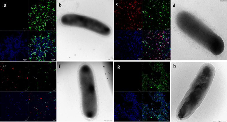
Results: In this study, we assess three approaches for the surface display of a panel of heterologous and homologous mature lipoproteins in E. coli: four from Neisseria meningitidis and four from the host strain that are known to be localised in the inner leaflet of the outer membrane. Constructs were made carrying the sequences coding for eight mature lipoproteins, each fused to the delivery portion of three different systems: the autotransporter adhesin involved in diffuse adherence-I (AIDA-I) from enteropathogenic E. coli, the Lpp'OmpA chimaera and a truncated form of the ice nucleation protein (INP), InaK-NC (N-terminal domain fused with C-terminal one) from Pseudomonas syringae. In contrast to what was observed for the INP constructs, when fused to the AIDA-I or Lpp'OmpA, most of the mature lipoproteins were displayed on the bacterial surface both at 37 and 25 °C as demonstrated by FACS analysis, confocal and transmission electron microscopy.

Conclusions: To our knowledge this is the first study that compares surface display systems using a number of passenger proteins. We have shown that the experimental conditions, including the choice of the carrier protein and the growth temperature, play an important role in the translocation of mature lipoproteins onto the bacterial surface. Despite all the optimization steps performed with the InaK-NC anchor motif, surface exposure of the passenger proteins used in this study was not achieved. For our experimental conditions, Lpp'OmpA chimaera has proved to be an efficient surface display system for the homologous passenger proteins although cell lysis and phenotype heterogeneity were observed. Finally, AIDA-I was found to be the best surface display system for mature lipoproteins (especially heterologous ones) in the E. coli host strain with no inhibition of growth and only limited phenotype heterogeneity.

Keywords: AIDA-I; Escherichia coli; Ice nucleation protein (InaK-NC); Lipoproteins; Lpp’OmpA chimaera; Surface display systems.


Conflict of interest statement

This work was sponsored by GlaxoSmithKline Biologicals SA. All authors have declared the following interests: MG, FG, LP, ID, DM and CB are employees of the GSK group of companies. CLG is a consultant for GSK, Italy. SN is a PhD student (University of Bologna) at GSK, Italy.

Figures

Fig. 1
Fig. 1
Schematic representation of the expression and display delivery systems. a Schematic representation of the domains of the constructs expressing recombinant versions of the eight proteins in the four different genetically engineered systems. b The panel illustrates schematically the display on the OM of each construct including 1) the full-length lipoprotein with its own signal peptide including lipobox lacking a carrier protein system and the mature portion of the lipoprotein fused to 2) AIDA-I, 3) Lpp’OmpA and 4) INP, InaK (N-domain + C-domain). In both panels (a and b), each protein domain is displayed with different colours. SP signal peptide, LP lipoprotein. OM outer membrane
Fig. 2
Fig. 2
Expression and surface display of the full-length lipoproteins in E. coli, at 37 °C. a Western blot of whole-cell lysates showing full-length lipoproteins. Lane 1: Marker, Lane 2: NmCsgG, Lane 3: NmMtrC, Lane 4: NmBamE, Lane 5: Nm putative lipoprotein, Lane 6: EcBamE, Lane 7: EcLolB, Lane 8: EcLptE and Lane 9: EcPal. FLAG-tag specific antibodies were used for detection. b, c FACS analysis of E. coli expressing full-length lipoproteins at 37 °C. At 37 °C, E. coli BL21DE3 (pET15b) expressing N. meningitidis lipoproteins: CsgG, MtrC, BamE and a putative lipoprotein (b) and E. coli lipoproteins (c): BamE, LolB, LptE and Pal were incubated with the monoclonal anti-FLAG antibody. The grey areas represent the fluorescence signals obtained with the control (BL21DE3-pET15b ∅). The coloured lines represent the full-length lipoproteins. Panel B (heterologous lipoproteins): Purple: CsgG, Dark Red: MtrC, Light Blue: nmBamE, Dark Green: putative lipoprotein; Panel C (homologous lipoproteins): Dark Blue: BamE, Light Green: LolB, Orange: LptE, Light Red: Pal
Fig. 3
Fig. 3
Expression and surface display of AIDA-I fusion proteins in E. coli, at 37 and 25 °C. Western blot of whole-cell lysates showing AIDA-I fusions proteins. Lane 1: Marker, Lane 2: NmCsgG, Lane 3: NmMtrC, Lane 4: NmBamE, Lane 5: Nm putative lipoprotein, Lane 6: EcBamE, Lane 7: EcLolB, Lane 8: EcLptE and Lane 9: EcPal, at 37 °C (a) and at 25 °C (b). FLAG-tag specific antibodies were used for detection. FACS analysis of E. coli BL21DE3 (pET15b) expressing AIDA-I fused to the N. meningitidis lipoproteins: CsgG, MtrC, BamE and putative lipoprotein at 37 °C (c) and 25 °C (d) and the E. coli lipoproteins: LolB, LptE, Pal and BamE at 37 °C (e) and 25 °C (f) were incubated with monoclonal anti-FLAG antibody. The grey areas represent the fluorescence signals obtained with the control (BL21DE3-pET15b ∅). The coloured lines represent the fused forms of the lipoproteins. Panels C and D (heterologous lipoproteins): Purple: CsgG, Dark Red: MtrC, Light Blue: nmBamE, Dark Green: putative lipoprotein; Panels E and F (homologous lipoproteins): Dark Blue: BamE, Light Green: LolB, Orange: LptE, Light Red: Pal
Fig. 4
Fig. 4
Expression and surface display of Lpp’OmpA fusion proteins in E. coli, at 37 and 25 °C. Western blot of whole-cell lysates showing Lpp’OmpA fusions proteins. Lane 1: Marker, Lane 2: Nm putative lipoprotein, Lane 3: NmMtrC, Lane 4: NmBamE, Lane 5: NmCsgG, Lane 6: EcBamE, Lane 7: EcLolB, Lane 8: EcLptE and Lane 9: EcPal, at 37 °C (a) and at 25 °C (b). FLAG-tag specific antibodies were used for detection. FACS analysis of E. coli BL21DE3 (pET15b) expressing Lpp’OmpA fused the N. meningitidis lipoproteins: CsgG, MtrC, BamE and putative lipoprotein, at 37 °C (c) and 25 °C (d) and the E. coli lipoproteins: LolB, LptE, Pal and BamE at 37 °C (e) and 25 °C (f) were incubated with monoclonal anti-FLAG antibody. The grey areas represent the fluorescence signals obtained with the control (BL21DE3-pET15b ∅). The coloured lines represent the fused forms of the lipoproteins. Panels C and D (heterologous lipoproteins): Purple: CsgG, Dark Red: MtrC, Light Blue: nmBamE, Dark Green: putative lipoprotein; Panels E and F (homologous lipoproteins): Dark Blue: BamE, Light Green: LolB, Orange: LptE, Light Red: Pal
Fig. 5
Fig. 5
Expression and surface display of InaK fusion proteins in E. coli, at 18 °C. Western blot of whole-cell lysates showing InaK fusions proteins. Lane 1: Marker; Lane 2: T7ExpressIq expressing InaK-NmBamE,at 18 °C (a). FACS analysis of E. coli T7ExpressIq (pET15b) expressing InaK fused the N. meningitidis lipoprotein BamE at 18 °C (b) was incubated with polyclonal anti-NmBamE antibodies. The grey area represents the fluorescence signal obtained with the control (T7ExpressIq pET15b ∅). The light blue coloured line represents the fused form of the NmBamE lipoprotein
Fig. 6
Fig. 6
Confocal and Transmission electron microscopy of BamE expressed in E. coli. E. coli BL21(DE3) expressing the full-length neisserial BamE (a and b), or fused to AIDA-I (c and d), or the E. coli BamE fused to Lpp’OmpA (e and f) grown at 25 °C, were incubated first with anti-FLAG antibodies while E. coli T7ExpressIq (pET15b) expressing InaK fused to the neisserial BamE grown at 18 °C, was incubated first with polyclonal anti-NmBamE antibodies (g and h). Subsequently samples were incubated with the secondary anti-mouse immunoglobulin G (whole molecule) Alexa fluor 568-conjugated. The lipoproteins can be visualized in red, the DNA in blue (DAPI) and the membranes in green (oregon green) (a, c, e and f). In transmission electron microscopy using immunogold labelling, the same samples were incubated with the secondary anti-mouse immunoglobulin G conjugated with 5 nm gold particles (b, d, f and h)

Similar articles

Cited by

References

    1. Gal I, Schlesinger O, Amir L, Alfonta L. Yeast surface display of dehydrogenases in microbial fuel-cells. Bioelectrochemistry. 2016;112:53–60. doi: 10.1016/j.bioelechem.2016.07.006. - DOI - PubMed
    1. Mathew E, Zhu H, Connelly SM, Sullivan MA, Brewer MG, Piepenbrink MS, et al. Display of the HIV envelope protein at the yeast cell surface for immunogen development. PLoS ONE. 2018;13(10):e0205756. doi: 10.1371/journal.pone.0205756. - DOI - PMC - PubMed
    1. Shang Y, Tesar D, Hötzel I. Modular protein expression by RNA trans-splicing enables flexible expression of antibody formats in mammalian cells from a dual-host phage display vector. Protein Eng Des Sel. 2015;28:437–444. doi: 10.1093/protein/gzv018. - DOI - PubMed
    1. Parola C, Mason DM, Zingg A, Neumeier D, Reddy ST. Genome engineering of hybridomas to generate stable cell lines for antibody expression. Methods Mol. Biol. 2018 - PubMed
    1. Nika L, Wallner J, Palmberger D, Koczka K, Vorauer-Uhl K, Grabherr R. Expression of full-length HER2 protein in Sf9 insect cells and its presentation on the surface of budded virus-like particles. Protein Expr Purif. 2017;136:27–38. doi: 10.1016/j.pep.2017.06.005. - DOI - PubMed

Publication types

MeSH terms