Observational Study
. 2021 Feb 2;11(1):2748.
doi: 10.1038/s41598-021-82536-2.

Central serous chorioretinopathy in active endogenous Cushing's syndrome

Affiliations
Observational Study

Central serous chorioretinopathy in active endogenous Cushing's syndrome

Joost Brinks et al. Sci Rep. .

Abstract

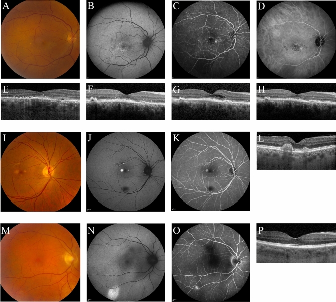
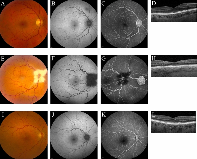
Multiple case series have provided evidence for a relatively high incidence of central serous chorioretinopathy (CSC) in patients with active Cushing's syndrome (CS). We describe the ophthalmological status in detail of consecutive patients with active endogenous CS (either de novo or recurrent active endogenous CS) in this prospective cohort study. All patients underwent complete ophthalmological examination, including multimodal imaging, which was performed shortly after establishing the diagnosis of active CS in hypercortisolemic state. Eleven CS patients (4 men, 7 women) with active hypercortisolism were included. Abnormalities reminiscent of (subclinical) CSC were found in 3 patients. Optical coherence tomography (OCT) revealed macular subretinal fluid in 1 patient, who was diagnosed as having active CSC and was successfully treated with half-dose photodynamic therapy. Two other patients showed CSC-like abnormalities: an unilateral pseudovitelliform lesion on OCT and hyperfluorescent changes on fluorescein angiography in one patient, and unilateral leakage on fluorescein angiography in the other patient. Mean subfoveal choroidal thickness on enhanced depth imaging OCT was 270 ± 40 μm (range, 178 - 357 μm). Retinal abnormalities resembling (subclinical) CSC may be more common than previously thought in patients with active CS, and may exist even in patients without visual complaints. Clinicians should have a low threshold for ophthalmological evaluation in case of a CS patient with visual symptoms since there may be therapeutic opportunities to prevent vision loss.


Conflict of interest statement

The authors declare no competing interests.

Figures

Figure 1
Figure 1
Multimodal imaging (AE) of the right eye of a 66-year-old female patient with active Cushing’s syndrome. The optical coherence tomography (OCT) scan (E) revealed a small amount of subretinal fluid (SRF) with hyperreflective debris. Fundus autofluorescence (B) mainly showed hyperautofluorescent changes macularly. On the mid-phase fluorescein angiography (C) 2 ‘hot spots’ of leakage were observed, nasally and temporally to the fovea. Mid-phase indocyanine green angiography (D) at that time did not provide evidence for a neovascularization. In the left eye of this patient, no abnormalities were detected on multimodal imaging (not shown). As SRF persisted after 6 months of leuprorelin treatment for Cushing’s syndrome, half-dose photodynamic therapy was performed, after which the SRF almost completely resolved (F). However, 3 months later intraretinal fluid occurred (G), and intravitreal injections with bevacizumab were scheduled. After 3 initial injections once per month, a treat-and-extend protocol was used. Eight weeks after the last bevacizumab injection until to date, SRF and intraretinal fluid on OCT had disappeared (H). Unfortunately, visual acuity did not improve. Multimodal imaging of the right eye of a 63-year-old male with active Cushing’s syndrome (IM). The foveal OCT scan (L) showed retinal pigment epithelium  alterations and parafoveal pseudovitelliform lesions. Corresponding hyperautofluorescent abnormalities were observed on fundus autofluorescence (J) and mid-phase fluorescein angiography (K). The abnormalities observed on multimodal imaging showed to be stable during follow-up, as can be seen on the OCT scan that was obtained at the last follow-up visit at 12 months after baseline (M). No abnormalities were observed in the left eye of this patient (imaging not shown). Multimodal imaging (NQ) of the right eye of a 61-year-old male with active Cushing’s syndrome. No abnormalities were seen on the OCT scan (Q). Mid-phase fluorescein angiography (P) showed a ‘hot spot’ of focal leakage, with an eccentric location outside the vascular arcade, and on fundus autofluorescence (O) hyperautofluorescent changes were observed in the same area. Unfortunately, this area was outside the covering area of the OCT scan, so possible presence of SRF could not be evaluated. No abnormalities were observed in the left eye of this patient (imaging not shown).
Figure 2
Figure 2
Multimodal imaging of ocular abnormalities not attributed to the spectrum of central serous chorioretinopathy. A 58-year-old male patient with Cushing’s syndrome due to a pituitary adenoma and a history of central retinal vein occlusion in the right eye (5 years ago) and grade 2 hypertensive retinopathy (3 years ago) showed mild parafoveal hyperfluorescent changes on fluorescein angiography (C). No abnormalities were observed on fundus photography (A), fundus autofluorescence imaging (B), and the foveal optical coherence tomography (OCT) scan (D). Furthermore, no abnormalities were found in the left eye on any of the imaging modalities (imaging not shown). The observed abnormalities in the right eye were stable after 1 year of follow-up after the first visit (imaging not shown). A 50-year-old male patient with Cushing’s syndrome due to a pituitary adenoma, who had a history of amblyopia in the right eye. Ophthalmological examination showed myelinated fibers which were present in both eyes (EG). Furthermore, on fluorescein angiography (G) congenital hypertrophy of the retinal pigment epithelium was visible in the right eye. Due to amblyopia in the right eye, an OCT scan of sufficient quality could not be obtained. Therefore, an OCT of the left eye is shown, on which no abnormalities were observed (H). Multimodal imaging of the right eye a 75-year-old female with Cushing’s syndrome due to an adrenal adenoma, which showed drusen on fundus photography (I), with only very mild changes on fundus autofluorescence imaging (J), mid-phase fluorescein angiography (K), and the foveal OCT scan (L). These observations were similar in the left eye (imaging not shown).

References

    1. Lindsay JR, Nansel T, Baid S, Gumowski J, Nieman LK. Long-term impaired quality of life in Cushing's syndrome despite initial improvement after surgical remission. J. Clin. Endocrinol. Metab. 2006;91:447–453. doi: 10.1210/jc.2005-1058. - DOI - PubMed
    1. van Haalen FM, Broersen LH, Jorgensen JO, Pereira AM, Dekkers OM. Management of endocrine disease: Mortality remains increased in Cushing's disease despite biochemical remission: a systematic review and meta-analysis. Eur. J. Endocrinol. 2015;172:143–149. doi: 10.1530/eje-14-0556. - DOI - PubMed
    1. Nieman LK. Cushing's syndrome: update on signs, symptoms and biochemical screening. Eur. J. Endocrinol. 2015;173:M33–38. doi: 10.1530/eje-15-0464. - DOI - PMC - PubMed
    1. Nieman LK, et al. The diagnosis of Cushing's syndrome: an Endocrine Society Clinical Practice Guideline. J. Clin. Endocrinol. Metab. 2008;93:1526–1540. doi: 10.1210/jc.2008-0125. - DOI - PMC - PubMed
    1. Daruich A, et al. Central serous chorioretinopathy: Recent findings and new physiopathology hypothesis. Prog. Retin. Eye Res. 2015;48:82–118. doi: 10.1016/j.preteyeres.2015.05.003. - DOI - PubMed

Publication types

MeSH terms