. 2021 Jan 26:16:147-158.
doi: 10.2147/COPD.S272711. eCollection 2021.

Rcn3 Suppression Was Responsible for Partial Relief of Emphysema as Shown by Specific Type II Alveolar Epithelial Cell Rcn3 CKO Mouse Model

Affiliations

Rcn3 Suppression Was Responsible for Partial Relief of Emphysema as Shown by Specific Type II Alveolar Epithelial Cell Rcn3 CKO Mouse Model

Qianyu Zhang et al. Int J Chron Obstruct Pulmon Dis. .

Abstract

Background: Chronic obstructive pulmonary disease (COPD), characterized by irreversible airflow limitation, is a highly prevalent lung disease worldwide and imposes increasing disease burdens globally. Emphysema is one of the primary pathological features contributing to the irreversible decline of pulmonary function in COPD patients, but the pathogenetic mechanisms remain unclear. Reticulocalbin 3 (Rcn3) is an endoplasmic reticulum (ER) lumen protein localized in the secretory pathway of living cells. Rcn3 in type II alveolar epithelial cell (AECIIs) has been reported to play a critical role in regulating perinatal lung development and bleomycin-induced lung injury-repair processes. We hypothesized that Rcn3 deficiency is associated with the development of emphysema during COPD, which is associated with the dysfunction of injury-repair modulated by alveolar epithelial cells.

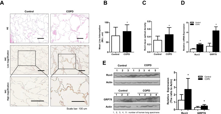
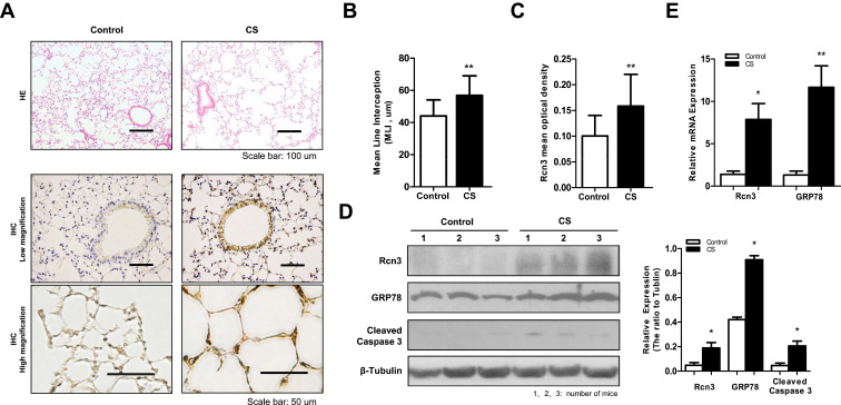
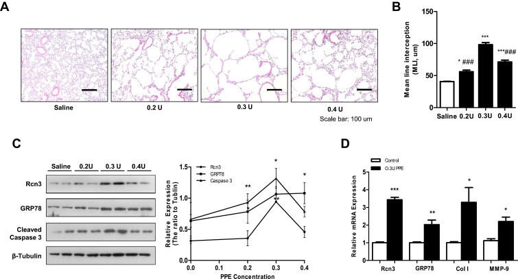
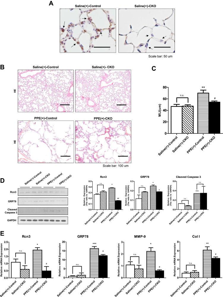
Materials and methods: We examined Rcn3 expression in lung specimens from COPD patients and non-COPD control patients undergoing lung lobectomy or pneumonectomy. Two mouse models of emphysema were established by cigarette smoke (CS) exposure and intratracheal instillation of porcine pancreatic elastase (PPE). Rcn3 expression was detected in the lung tissues from these mice. Furthermore, conditional knockout (CKO) mice with Rcn3 deletion specific to AECIIs were used to explore the role of Rcn3 in PPE-induced emphysema progression. Rcn3 protein expression in lung tissues was evaluated by Western blot and immunohistochemistry. Rcn3 mRNA expression in lung tissues was detected by qPCR.

Results: Rcn3 expression was significantly increased in the lung specimens from COPD patients versus non-COPD patients and the level of Rcn3 increase was associated with the degree of emphysema. Rcn3 expression were also significantly up-regulated in both CS-induced and PPE-induced emphysematous mouse lungs. Moreover, the selective ablation of Rcn3 in AECIIs significantly alleviated severity of the mouse emphysema in response to intratracheal installation of PPE.

Conclusion: Our data, for the first time, indicated that suppression of Rcn3 expression in AECIIs has a beneficial effect on PPE-induced emphysema.

Keywords: COPD; Rcn3; emphysema; type II alveolar epithelial cell.


Conflict of interest statement

The authors declare that they have no competing or conflicts of interest.

Figures

Figure 1
Figure 1
Rcn3 expression in lung tissues from COPD patients. A total of 70 specimens were divided into COPD group and Control group according to the pulmonary function test and pathological changes of emphysema. (A) Photomicrography of pulmonary parenchyma stained with H&E (upper panels). Bar size: 100 μm; IHC assays of Rcn3 from COPD group lungs and Control group lungs (low and high magnifications, lower panels). Bar size: 100 μm; (B) The MLI analysis of H&E stained lung sections (82.78 ± 9.417 μm vs.104.8 ± 5.789 μm, n = 21 in Control group and n = 34 in COPD group, p<0.05); (C) Quantification of the mean optical density of Rcn3 expression in the lung tissues according to the Rcn3 IHC assays (3 random views, 100× amplification for one subject, n = 11–12 per group, p<0.05); (D) Qualitative PCR analyses of the mRNA expressions of Rcn3 and GRP78; the data were normalized to the GAPDH content and were analyzed by the 2−ΔΔCt method relative to the Control group; n = 6 in Control group and n = 10 in COPD group; (E) The protein levels of Rcn3 and GRP78 were examined by Western blot analysis and the ratios to Actin presented by bar graph; n = 6 in Control group and n = 10 in COPD group. Data presented as mean ± SEM; *p<0.05 versus the Control group.
Figure 2
Figure 2
Rcn3 expression in lung tissues from CS-induced mouse model of emphysema. A total of 16 C57BL/6 mice (6 weeks old) were exposed to cigarette smoke for 6 months as emphysema mice (CS group) and 16 C57BL/6 mice were exposed to air for 6 months as Control mice (Control group). (A) Photomicrography of pulmonary parenchyma stained with H&E (upper panels). Bar size: 100 μm; IHC assays of Rcn3 (low and high magnifications, lower panels). Bar size: 50 μm; (B) Morphometric analysis of the MLI ((44.00 ± 2.528 μm vs.56.81 ± 3.053 μm, n = 16 per group, p<0.01); (C) The mean optical density of Rcn3 expression was measured in the lung tissues according to the Rcn3 IHC assays (3 random views, 100× amplification for one subject, n = 16 per group, p<0.01); (D) Representative WB for protein levels of Rcn3, GRP78 and Cleaved caspase-3 in lungs and the ratios to β-Tubulin presented by bar graph, n = 8 per group; (E) Expressions of Rcn3 and GRP78 mRNA were determined with Qualitative PCR analysis, n = 9–10 per group. Data presented as mean ± SEM; *p<0.05, **p<0.01 versus the Control group.
Figure 3
Figure 3
Rcn3 expression in lung tissues from the elastase-induced mouse model of emphysema. (A) Lungs after saline or PPE instillation at 0.2 U, 0.3 U and 0.4 U/per mouse were compared in H&E-stained tissue sections. Bar size: 100 μm; (B) The MLI analysis of lung sections after saline or PPE instillation (40.58 ± 0.53 μm, 56.10 ± 2.45 μm, 98.23 ± 3.21 μm and 71.22 ± 2.84 μm, respectively, n = 5 per group); (C) The PPE instillation induced the protein expressions of Rcn3, GRP78, Cleaved caspase-3, and the ratios to β-Tubulin expression are represented by graphs, n = 5 per group; (D) Qualitative PCR analysis of the mRNA expressions of Rcn3, GRP78, Collagen I (Col (I) and MMP-9 in the lungs at 4 weeks after PPE (0.3 U/per mouse) or saline treatment, n = 5 per group. Data presented as mean ± SEM; *p<0.05, **p<0.01, ***p<0.001, versus the Saline group, ###p<0.001, versus the 0.3 U PPE group.
Figure 4
Figure 4
The selective deletion of Rcn3 in AECIIs alleviated elastase-induced emphysema. (A) IHC assays of Rcn3 from lungs in saline-treated WT mice or saline-treated CKO mice (arrows indicate the corners of alveoli, suggestive of AECIIs). Bar size: 50 μm; (B) H&E staining of lung tissues from mice in Saline(+)-Control, Saline(+)-CKO, PPE(+)-Control and PPE(+)-CKO groups. Bar size: 100 μm; (C) The MLI analysis of lung sections from the four groups of mice (46.85 ± 3.56 μm, 47.45 ± 2.268 μm, 70.07 ± 5.067 μm, and 54.34 ± 1.977 μm, respectively, n = 9–10 per group); (D) The protein levels of Rcn3, GRP78 and Cleaved caspase-3 in Saline(+)-Control, Saline(+)-CKO, PPE(+)-Control, and PPE(+)-CKO mice were examined by Western blot analysis and the ratios to GAPDH presented by bar graph, n = 5 per group; (E) Qualitative PCR analysis of the mRNA expressions of Rcn3, GRP78, MMP-9, and Collagen I in Saline(+)-Control, Saline(+)-CKO, PPE(+)-Control, and PPE(+)-CKO mice, n = 5 per group. Data presented as mean ± SEM; *p<0.05, **p<0.01, ***p<0.001, versus Saline(+)-Control group; #p<0.05, ##p<0.01, versus PPE(+)-Control group.

References

    1. Singh D, Agusti A, Anzueto A, et al. Global strategy for the diagnosis, management, and prevention of chronic obstructive lung disease. GOLD Science Committee Report 2019. 2019;53. - PubMed
    1. Adeloye D, Chua S, Lee C, et al. Global and regional estimates of COPD prevalence. Systematic Review Meta-Analysis. 2015;5:020415. - PMC - PubMed
    1. GBD 2013 Mortality and Causes of Death Collaborators. Global, regional, and national age-sex specific all-cause and cause-specific mortality for 240 causes of death, 1990–2013: a systematic analysis for the global burden of disease study 2013. J Lancet:. 2015;385:117–171. doi:10.1016/S0140-6736(14)61682-2 - DOI - PMC - PubMed
    1. Miniño A, Xu J. National vital statistics reports: from the centers for disease, prevention national center for health statistics, national vital statistics system: deaths: preliminary data for 2008. Control Kochanek Kd. 2010;59:1–52. - PubMed
    1. Vogelmeier C, Criner G, Martínez F, et al. Global strategy for the diagnosis, management, and prevention of chronic obstructive lung disease 2017 report. GOLD Executive Summary. 2017;53:128–149. - PubMed

Substances