. 2021 Jan 16:2021:6638575.
doi: 10.1155/2021/6638575. eCollection 2021.

IL-1 Receptor Antagonist Protects the Osteogenesis Capability of Gingival-Derived Stem/Progenitor Cells under Inflammatory Microenvironment Induced by Porphyromonas gingivalis Lipopolysaccharides

Affiliations

IL-1 Receptor Antagonist Protects the Osteogenesis Capability of Gingival-Derived Stem/Progenitor Cells under Inflammatory Microenvironment Induced by Porphyromonas gingivalis Lipopolysaccharides

Yuxin Zhao et al. Stem Cells Int. .

Abstract

Mesenchymal stem cells (MSCs) have been considered to be a future treatment option for periodontitis due to their excellent regenerative capability. However, it is still a challenge to protect MSCs' biological properties from multiple bacterial toxins in local inflammatory environment. The present study is aimed at investigating the treatment effect of interleukin-1 receptor antagonist (IL-1ra) on cell proliferation, migration, and osteogenic differentiation of gingival-derived mesenchymal stem cells (GMSCs) under an inflammatory microenvironment induced by Porphyromonas gingivalis lipopolysaccharides (P. gingivalis-LPS). GMSCs derived from Sprague-Dawley (SD) rats' free gingival tissues were treated with P. gingivalis-LPS (10 μg/mL) to create in vitro inflammatory environment. Different concentrations of IL-1ra (0.01-1 μg/mL) were used to antagonize the negative effect of LPS. Cell behaviors including proliferation, cloning formation unit (CFU), cell migration, osteogenic differentiation, mineral deposition, and cytokine production were assessed to investigate the protection effect of IL-1ra on GMSCs under inflammation. The toll-like receptor 4 (TLR4)/nuclear factor kappa B (NF-κB) pathway activated by LPS was evaluated by real-time quantitative polymerase chain reaction (RT-PCR) and western blot. In response to P. gingivalis-LPS treatment, cell numbers, cloning formation rate, cell migration rate, proinflammatory cytokine production, and osteogenic differentiation-associated protein/mRNA expressions as well as mineralized nodules were suppressed in a time-dependent manner. These negative effects were effectively attenuated by IL-1ra administration in a time- and dose-dependent manner. In addition, mRNA expressions of TLR4 and IkBα decreased dramatically when IL-1ra was added into LPS-induced medium. IL-1ra also reversed the LPS-induced TLR4/NF-κB activation as indicated by western blot. The present study revealed that IL-1ra decreased inflammatory cytokine production in a supernatant, so as to protect GMSCs' osteogenesis capacity and other biological properties under P. gingivalis-LPS-induced inflammatory environment. This might be explained by IL-1ra downregulating TLR4-mediated NF-κB signaling pathway activation.


Conflict of interest statement

The authors declare no conflict of interest regarding the publication of this article.

Figures

Figure 1
Figure 1
Characterization and multidirectional differentiation potential of stem/progenitor cells from rat gingival tissues. (a) Microscopic appearance of rGMSC morphology (20x). (b) Osteogenic differentiation of rGMSCs after 21 days of osteogenic induction. Positive staining of calcified deposits is observed (200x). (c) Adipogenic differentiation of rGMSCs after 21 days of adipogenic induction. Positive staining of lipid droplets is observed (200x). (d) Chondrogenic differentiation of rGMSCs after 21 days of chondrogenic induction. Positive staining of glycosaminoglycans is observed (200x).
Figure 2
Figure 2
FACS identification of the surface marker expressions of rGMSCs. CD73, CD90, CD105, CD146, and Stro-1 were highly expressed, while rGMSCs were negative for CD14, CD34, and CD45.
Figure 3
Figure 3
Colony formation of rGMSCs under inflammatory and anti-inflammatory environment. GIEMSA staining results for the colony-forming unit (CFU) assay (G1: GMSCs; G2: GMSCs+LPS; G3: GMSCs+LPS+10 ng/mL IL-1ra; G4: GMSCs+LPS+100 ng/mL IL-1ra; and G5: GMSCs+LPS+1 μg/mL IL-1ra).
Figure 4
Figure 4
Effect of P. gingivalis-LPS and IL-1ra on rGMSC colony formation efficiency at 14 days. ∗∗P < 0.01, ∗∗∗P < 0.001 vs. GMSCs; #P < 0.05, ###P < 0.001 vs. GMSCs+LPS.
Figure 5
Figure 5
Effect of P. gingivalis-LPS and IL-1ra on rGMSC proliferation potential at 1, 3, and 7 days. P < 0.05, ∗∗P < 0.01 vs. GMSCs; #P < 0.05, ##P < 0.01 vs. GMSCs+LPS.
Figure 6
Figure 6
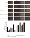
Effect of P. gingivalis-LPS and IL-1ra on rGMSC migration capacity at 24, 48, and 72 hours. (a) Migration of rGMSCs to cover the scratch area. (b) Cell migration rate in the control group, inflammatory group, and anti-inflammatory groups at different time points. P < 0.05 vs. GMSCs; #P < 0.05 vs. GMSCs+LPS.
Figure 7
Figure 7
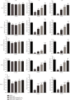
mRNA expression of osteogenic genes including ALP, COL-I, OPN, OCN, and RNX2 in different groups after 24 hours, 7 days, and 14 days of osteogenic induction, detected by RT-PCR. P < 0.05, ∗∗P < 0.01, and ∗∗∗P < 0.001 vs. GMSCs; #P < 0.05, ##P < 0.01, and ###P < 0.001 vs. GMSCs+LPS.
Figure 8
Figure 8
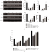
rGMSC osteogenic gene expression on protein level and calcified nodule deposition. (a) Expression of osteogenic-related proteins ALP and OCN in rGMSCs after 7 days and 14 days of osteogenic induction, detected by western blot (G1: GMSCs; G2: GMSCs+LPS; G3: GMSCs+LPS+10 ng/mL IL-1ra; G4: GMSCs+LPS+100 ng/mL IL-1ra; and G5: GMSCs+LPS+1 μg/mL IL-1ra). (b) Mineralized nodule formation by rGMSCs after 14 days, 21 days, and 28 days of osteogenic induction, detected by Alizarin Red staining. P < 0.05, ∗∗P < 0.01, and ∗∗∗P < 0.001 vs. GMSCs; #P < 0.05, ##P < 0.01, and ###P < 0.001 vs. GMSCs+LPS.
Figure 9
Figure 9
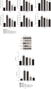
IL-1ra attenuates the P. gingivalis-LPS-activated TLR4-mediated NF-κB pathway in rGMSCs. RT-PCR and western blot analysis were performed. (a) mRNA expressions of TLR4 and IκBα were analyzed by RT-PCR at 1, 7, and 14 days. (b) Protein expressions of phosphorylated IκBα, IκBα, NF-κB phosphorylated p65, and NF-κB p65 after 1-hour induction in different groups (G1: GMSCs; G2: GMSCs+LPS; G3: GMSCs+LPS+10 ng/mL IL-1ra; G4: GMSCs+LPS+100 ng/mL IL-1ra; and G5: GMSCs+LPS+1 μg/mL IL-1ra). (c) The ratio of p-IκBα/IκBα and NF-κB p-p65/p65 in different groups. P < 0.05, ∗∗P < 0.01, and ∗∗∗P < 0.001 vs. GMSCs; #P < 0.05, ##P < 0.01, and ###P < 0.001 vs. GMSCs+LPS.
Figure 10
Figure 10
Effects of P. gingivalis-LPS and IL-1ra on cytokine release in rGMSCs after 12, 24, and 48 hours, as measured by using ELISA kits. (a) The production of IL-1β. (b) The production of TNF-α. (c) The production of IL-1ra. P < 0.05, ∗∗P < 0.01 vs. GMSCs; #P < 0.05, ##P < 0.01 vs. GMSCs+LPS.

Similar articles

Cited by

References

    1. Mysak J., Podzimek S., Sommerova P., et al. Porphyromonas gingivalis: major periodontopathic pathogen overview. Journal of immunology research. 2014;2014:8.476068 - PMC - PubMed
    1. Cheng R., Liu W., Zhang R., Feng Y., Bhowmick N. A., Hu T. Porphyromonas gingivalis-derived lipopolysaccharide combines hypoxia to induce caspase-1 activation in periodontitis. Frontiers in cellular and infection microbiology. 2017;7:p. 474. - PMC - PubMed
    1. Bostanci N., Belibasakis G. N. Porphyromonas gingivalis: an invasive and evasive opportunistic oral pathogen. FEMS Microbiology Letters. 2012;333(1):1–9. doi: 10.1111/j.1574-6968.2012.02579.x. - DOI - PubMed
    1. Liu A. Q., Hu C. H., Jin F., Zhang L. S., Xuan K. Contributions of bioactive molecules in stem cell-based periodontal regeneration. International journal of molecular sciences. 2018;19(4) - PMC - PubMed
    1. Kuo T. F., Lee S. Y., Wu H. D., Poma M., Wu Y. W., Yang J. C. An in vivo swine study for xeno-grafts of calcium sulfate-based bone grafts with human dental pulp stem cells (hDPSCs) Materials for biological applications. 2015;50:19–23. doi: 10.1016/j.msec.2015.01.092. - DOI - PubMed

LinkOut - more resources