

Giant Tuberculomas of Brain: Rare Neoplastic Mimic
- PMID: 33531933
- PMCID: PMC7847125
- DOI: 10.4103/jpn.JPN_78_19
Giant Tuberculomas of Brain: Rare Neoplastic Mimic
Abstract
Objective: Tuberculosis continues to be a major infectious disease in developing parts of the world. Primarily central nervous system tuberculosis manifests as meningitis, tuberculoma, or a brain abscess; however, rarely it may manifest as a large neoplastic mass such as lesion known as giant tuberculoma. Especially in central parts of India, the incidence of giant tuberculoma is quite high in pediatric population that too in posterior fossa of brain. Often, they are wrongly reported as neoplastic masses on imaging. The objective of this study was to evaluate different imaging appearances of a giant tuberculoma.
Materials and methods: In this prospective study, all cases of giant tuberculoma presenting to a large tertiary care center in central India for 2 years (duration 2016-2018) were imaged and followed up. A total of nine patients, six females and three males, aged 4-16 years were studied on a 3-Tesla Siemens magnetic resonance imaging (MRI) scanner.
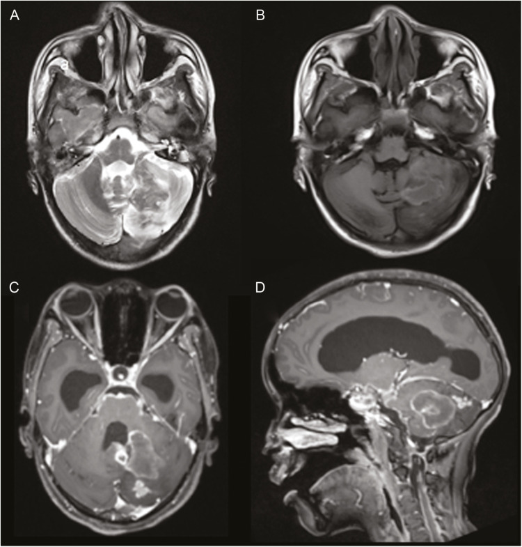
Results: In total, nine patients were included with 11 giant tuberculomas. Of 11, eight were infratentorial and three were supratentorial in location. On T2-weighted image sequence, these lesions showed central hypointensity with a peripheral hyperintense rim. Most observed finding on T1-weighted image sequence was central isointensity with peripheral hyperintense rim. Advanced imaging sequences such as magnetic resonance spectroscopy and magnetization transfer were also applied.
Conclusion: To the best of our knowledge, this is the largest series of giant tuberculoma in the pediatric population reported so far in any part of the world. We have described the various MRI imaging findings of this lesion in great details. Management of such rare cases and pertinent literature is reviewed briefly.
Keywords: Giant tuberculoma; neoplastic mimicker; pediatric population; posterior fossa.
Copyright: © 2020 Journal of Pediatric Neurosciences.
Conflict of interest statement
There are no conflicts of interest.
Figures


References
-
- Mukherjee S, Das R, Begum S. Tuberculoma of the brain: a diagnostic dilemma: magnetic resonance spectroscopy a new ray of hope. J Assoc Chest Physicians. 2015;3:3–8.
-
- Simsek H, Kutiay M, Colak A, Haholu A, Kaya H, Ozyurt M. Mehmet Nusret DEMIRCAN concomitant tubercular and fungal cerebellar abscess in an immunocompromised girl. Turkish Neurosurg. 2013;23:88–94. - PubMed
-
- Saxena S, Prakash M, Kumar S, Gupta RK. Comparative evaluation of magnetization transfer contrast and fluid attenuated inversion recovery sequences in brain tuberculoma. Clin Radiol. 2005;60:787–93. - PubMed
-
- Chakraborty AK. Prevalence and incidence of tuberculosis infection and disease in India: a comprehensive review WHO/TB/97.231. Geneva, Switzerland: World Health Organization; 1997. pp. 1–26.
LinkOut - more resources
Full Text Sources