

Radiomics in Stroke Neuroimaging: Techniques, Applications, and Challenges
- PMID: 33532134
- PMCID: PMC7801280
- DOI: 10.14336/AD.2020.0421
Radiomics in Stroke Neuroimaging: Techniques, Applications, and Challenges
Abstract
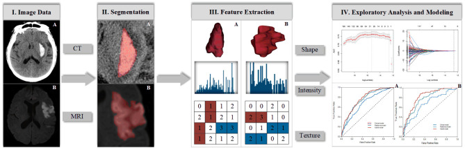
Stroke is a leading cause of disability and mortality worldwide, resulting in substantial economic costs for post-stroke care each year. Neuroimaging, such as cranial computed tomography or magnetic resonance imaging, is the backbone of stroke management strategies, which can guide treatment decision-making (thrombolysis or hemostasis) at an early stage. With advances in computational technologies, particularly in machine learning, visual image information can now be converted into numerous quantitative features in an objective, repeatable, and high-throughput manner, in a process known as radiomics. Radiomics is mainly used in the field of oncology, which remains an area of active research. Over the past few years, investigators have attempted to apply radiomics to stroke in the hope of gaining benefits similar to those obtained in cancer management, i.e., in promoting the development of personalized precision medicine. Currently, radiomic analysis has shown promise for a variety of applications in stroke, including the diagnosis of stroke lesions, early prediction of outcomes, and evaluation for long-term prognosis. In this article, we elaborate the contributions of radiomics to stroke, as well as the subprocesses and techniques involved in radiomics studies. We also discuss the potential challenges facing its widespread implementation in routine practice and the directions for future research.
Keywords: decision-making; neuroimaging; radiomics; stroke; texture analysis.
copyright: © 2021 Chen et al.
Conflict of interest statement
Conflict of Interest The authors declare no conflict of interest.
Figures
References
-
- Rajsic S, Gothe H, Borba HH, Sroczynski G, Vujicic J, Toell T, et al. (2018). Economic burden of stroke: a systematic review on post-stroke care. The European Journal of Health Economics, 20:107-134. - PubMed
-
- Buckler AJ, Bresolin L, Dunnick NR, Sullivan DC, Group (2011). A collaborative enterprise for multi-stakeholder participation in the advancement of quantitative imaging. Radiology, 258:906-914. - PubMed
Publication types
LinkOut - more resources
Full Text Sources
Other Literature Sources
Medical
Miscellaneous