

Long-Term Safety Evaluation of Continuous Intraocular Delivery of Aflibercept by the Intravitreal Gene Therapy Candidate ADVM-022 in Nonhuman Primates
- PMID: 33532145
- PMCID: PMC7846953
- DOI: 10.1167/tvst.10.1.34
Long-Term Safety Evaluation of Continuous Intraocular Delivery of Aflibercept by the Intravitreal Gene Therapy Candidate ADVM-022 in Nonhuman Primates
Abstract
Purpose: To evaluate the long-term safety of vascular endothelial growth factor (VEGF) suppression with sustained aflibercept expression after a single intravitreal injection (IVI) of ADVM-022, an anti-VEGF gene therapy, in non-human primates (NHPs).
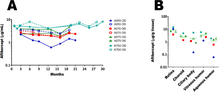
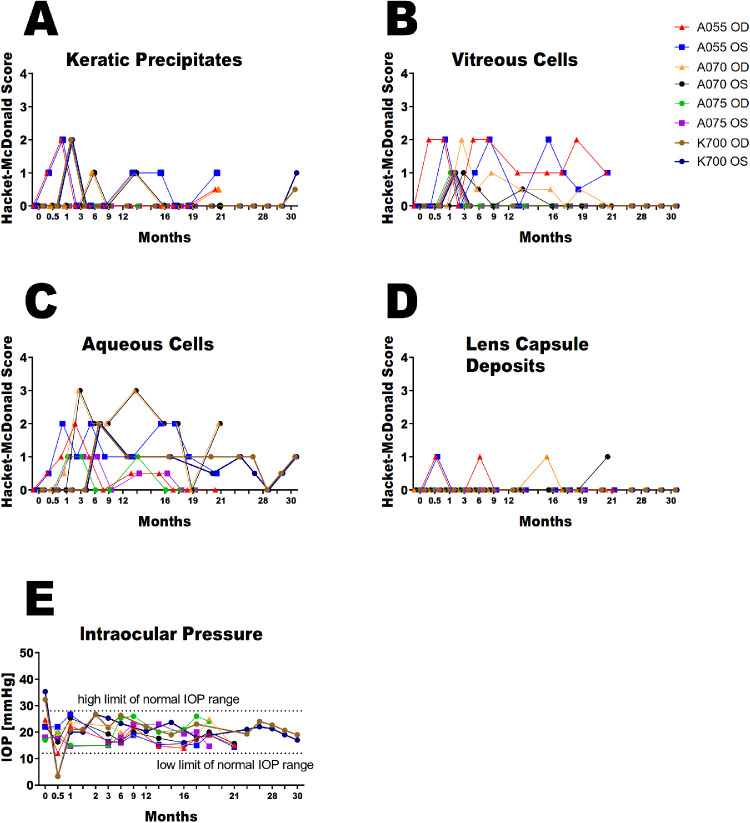
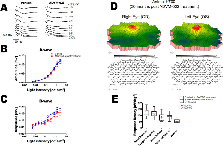
Methods: Non-human primates received bilateral IVI of ADVM-022, a gene therapy vector encoding aflibercept, a standard of care for the treatment of VEGF-based retinal disease. Aflibercept levels from ocular fluids and tissues were measured. Ocular inflammation was assessed by slit lamp biomicroscopy and fundoscopy. The integrity of the retinal structure was analyzed by optical coherence tomography and blue light fundus autofluorescence and electroretinography was performed to determine retinal function. Histologic evaluation of the retina was performed at the longest time point measured (2.5 years after injection).
Results: Sustained expression of aflibercept was noted out to the last time point evaluated. Mild to moderate inflammatory responses were observed, which trended toward spontaneous resolution without anti-inflammatory treatment. No abnormalities in retinal structure or function were observed, as measured by optical coherence tomography and electroretinography, respectively. RPE integrity was maintained throughout the study; no histologic abnormalities were observed 2.5 years after ADVM-022 IVI.
Conclusions: In non-human primates, long-term, sustained aflibercept expression and the resulting continuous VEGF suppression by a single IVI of ADVM-022, appears to be safe, with no measurable adverse effects on normal retinal structure and function evaluated out to 2.5 years.
Translational relevance: Together with the results from previous ADVM-022 preclinical studies, these data support the evaluation of this gene therapy candidate in clinical trials as a potential durable treatment for various VEGF-mediated ophthalmic disorders.
Keywords: ERG; OCT; adeno-associated viral vector; aflibercept; anti-VEGF.
Copyright 2021 The Authors.
Conflict of interest statement
Disclosure: S. Kiss, Adverum (C, R), Novartis (C, F), Optos (C, F), Genentech/Roche (C, F), Regeneron (C, F), Regenx Bio (R), Fortress Bio (R); K. Oresic Bender (E); R.N. Grishanin (E); K.M. Hanna (E); J.D. Nieves (E); P. Sharma (E); A.T. Nguyen (E); R.J. Rosario, None; J.S. Greengard (E); C.M. Gelfman (E); M. Gasmi (S)
Figures

Similar articles
-
Comprehensive Preclinical Assessment of ADVM-022, an Intravitreal Anti-VEGF Gene Therapy for the Treatment of Neovascular AMD and Diabetic Macular Edema.J Ocul Pharmacol Ther. 2021 Apr;37(3):181-190. doi: 10.1089/jop.2021.0001. J Ocul Pharmacol Ther. 2021. PMID: 33835848 Free PMC article.
-
Analysis of Aflibercept Expression in NHPs following Intravitreal Administration of ADVM-022, a Potential Gene Therapy for nAMD.Mol Ther Methods Clin Dev. 2020 Jun 12;18:345-353. doi: 10.1016/j.omtm.2020.06.007. eCollection 2020 Sep 11. Mol Ther Methods Clin Dev. 2020. PMID: 32671137 Free PMC article.
-
Preclinical Evaluation of ADVM-022, a Novel Gene Therapy Approach to Treating Wet Age-Related Macular Degeneration.Mol Ther. 2019 Jan 2;27(1):118-129. doi: 10.1016/j.ymthe.2018.11.003. Epub 2018 Nov 13. Mol Ther. 2019. PMID: 30528929 Free PMC article.
-
[Basic in vitro studies on VEGF inhibition with aflibercept: similarities and differences to other VEGF-binding therapeutic proteins].Klin Monbl Augenheilkd. 2015 Mar;232(3):295-302. doi: 10.1055/s-0034-1383142. Epub 2014 Nov 13. Klin Monbl Augenheilkd. 2015. PMID: 25393440 Review. German.
-
Same-Day Bilateral Intravitreal Anti-Vascular Endothelial Growth Factor Injections: Experience of a Large Canadian Retina Center.Ophthalmologica. 2019;242(1):1-7. doi: 10.1159/000499115. Epub 2019 Mar 29. Ophthalmologica. 2019. PMID: 30928983 Review.
Cited by
-
Anti-Scg3 Gene Therapy to Treat Choroidal Neovascularization in Mice.Biomedicines. 2023 Jul 6;11(7):1910. doi: 10.3390/biomedicines11071910. Biomedicines. 2023. PMID: 37509549 Free PMC article.
-
Review of gene therapies for age-related macular degeneration.Eye (Lond). 2022 Feb;36(2):303-311. doi: 10.1038/s41433-021-01842-1. Epub 2022 Jan 11. Eye (Lond). 2022. PMID: 35017696 Free PMC article. Review.
-
Lentiviral delivered aflibercept OXB-203 for treatment of neovascular AMD.Mol Ther Methods Clin Dev. 2023 Jul 15;30:350-366. doi: 10.1016/j.omtm.2023.07.001. eCollection 2023 Sep 14. Mol Ther Methods Clin Dev. 2023. PMID: 37637380 Free PMC article.
-
Inefficacy of anti-VEGF therapy reflected in VEGF-mediated photoreceptor degeneration.Mol Ther Nucleic Acids. 2024 Mar 25;35(2):102176. doi: 10.1016/j.omtn.2024.102176. eCollection 2024 Jun 11. Mol Ther Nucleic Acids. 2024. PMID: 38689803 Free PMC article.
-
Comprehensive Preclinical Assessment of ADVM-022, an Intravitreal Anti-VEGF Gene Therapy for the Treatment of Neovascular AMD and Diabetic Macular Edema.J Ocul Pharmacol Ther. 2021 Apr;37(3):181-190. doi: 10.1089/jop.2021.0001. J Ocul Pharmacol Ther. 2021. PMID: 33835848 Free PMC article.
References
-
- Parikh R, Pirakitikulr N, Chhablani J, Sakurada Y, Singh RP, Modi YS.. A multinational comparison of anti-vascular endothelial growth factor use: the United States, the United Kingdom, and Asia-Pacific. Ophthalmol Retina . 2019; 3(1): 16–26. - PubMed
-
- Rofagha S, Bhisitkul RB, Boyer DS, Sadda SR, Zhang K, Group S-US. Seven-year outcomes in ranibizumab-treated patients in ANCHOR, MARINA, and HORIZON: a multicenter cohort study (SEVEN-UP). Ophthalmology . 2013; 120(11): 2292–2299. - PubMed
-
- Ciulla TA, Hussain RM, Pollack JS, Williams DF.. Visual acuity outcomes and anti-vascular endothelial growth factor therapy intensity in neovascular age-related macular degeneration patients: a real-world analysis of 49 485 eyes. Ophthalmol Retina . 2020; 4(1): 19–30. - PubMed
Publication types
MeSH terms
Substances
LinkOut - more resources
Full Text Sources
Other Literature Sources
