. 2021 Jan;11(1):71-88.
doi: 10.1016/j.apsb.2020.09.006. Epub 2020 Sep 18.

Activated PKB/GSK-3 β synergizes with PKC- δ signaling in attenuating myocardial ischemia/reperfusion injury via potentiation of NRF2 activity: Therapeutic efficacy of dihydrotanshinone-I

Affiliations

Activated PKB/GSK-3 β synergizes with PKC- δ signaling in attenuating myocardial ischemia/reperfusion injury via potentiation of NRF2 activity: Therapeutic efficacy of dihydrotanshinone-I

Hao Zeng et al. Acta Pharm Sin B. 2021 Jan.

Abstract

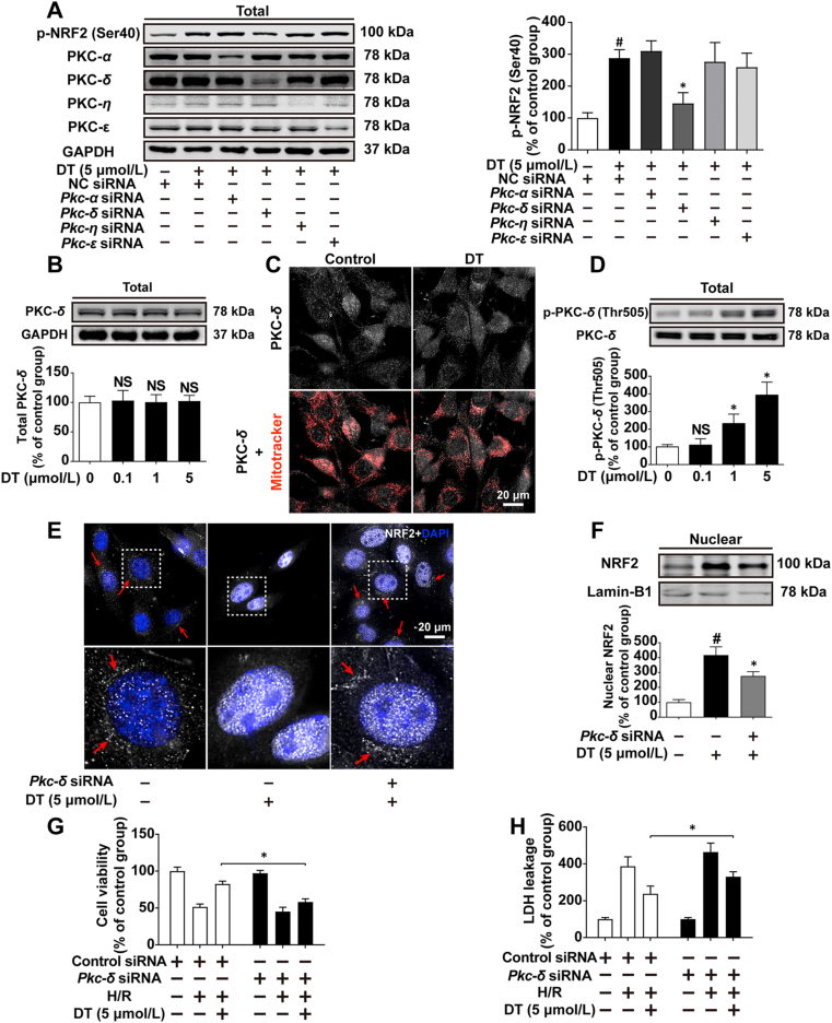
Disrupted redox status primarily contributes to myocardial ischemia/reperfusion injury (MIRI). NRF2, the endogenous antioxidant regulator, might provide therapeutic benefits. Dihydrotanshinone-I (DT) is an active component in Salvia miltiorrhiza with NRF2 induction potency. This study seeks to validate functional links between NRF2 and cardioprotection of DT and to investigate the molecular mechanism particularly emphasizing on NRF2 cytoplasmic/nuclear translocation. DT potently induced NRF2 nuclear accumulation, ameliorating post-reperfusion injuries via redox alterations. Abrogated cardioprotection in NRF2-deficient mice and cardiomyocytes strongly supports NRF2-dependent cardioprotection of DT. Mechanistically, DT phosphorylated NRF2 at Ser40, rendering its nuclear-import by dissociating from KEAP1 and inhibiting degradation. Importantly, we identified PKC-δ-(Thr505) phosphorylation as primary upstream event triggering NRF2-(Ser40) phosphorylation. Knockdown of PKC-δ dramatically retained NRF2 in cytoplasm, convincing its pivotal role in mediating NRF2 nuclear-import. NRF2 activity was further enhanced by activated PKB/GSK-3β signaling via nuclear-export signal blockage independent of PKC-δ activation. By demonstrating independent modulation of PKC-δ and PKB/GSK-3β/Fyn signaling, we highlight the ability of DT to exploit both nuclear import and export regulation of NRF2 in treating reperfusion injury harboring redox homeostasis alterations. Coactivation of PKC and PKB phenocopied cardioprotection of DT in vitro and in vivo, further supporting the potential applicability of this rationale.

Keywords: Cytoplasmic/nuclear translocation; Dihydrotanshinone I; Ischemia/reperfusion injury; NRF2; PKB/GSK-3β/Fyn; PKC-δ; Phosphorylation; Redox homeostasis.


Figures

Image 1
Graphical abstract
Figure 1
Figure 1
DT induces NRF2 activation and ameliorates myocardial ischemia/reperfusion and hypoxia/reoxygenation injury with maintained redox homeostasis. (A) Chemical structure of dihydrotanshinone I. (B) Representative images and calculated infarct size of the myocardium, scale bar = 2 mm (n = 5). (C) Echocardiogram of LV and the calculated EF and FS (n = 6). (D) Representative images of WGA-stained LV sections and the calculated cross-sectional area of cardiomyocytes, scale bar = 25 μm (n = 6). (E) Representative images of 8-OHDG/α-actinin/DAPI-stained LV sections and the calculated nuclear 8-OHDG intensity, scale bar = 100 μm (n = 6). (F) Nuclear protein level of NRF2 in the myocardium of sham, I/R and DT-treated mice (n = 5). (G)–(I) Cell viability, extracellular LDH leakage and intracellular MDA content of cardiomyocytes (n = 5). (J) Representative images of CELLROX-stained cardiomyocytes, scale bar = 20 μm (n = 5). (K) Nuclear protein level of NRF2 in control, H/R and DT-treated cardiomyocytes (n = 5). Data are expressed as mean ± SD, #P < 0.05 versus sham or control group; ∗P < 0.05 versus I/R or H/R group.
Figure 2
Figure 2
Genetically knockout of Nrf2 abolishes the cardioprotective effects of DT against MIRI. (A) and (B) Protein level and gene expression of Nrf2 in cardiac tissues of wild type (WT) and Nrf2-KO mice (n = 5). (C)–(E) Gene expression of Ho-1, Nqo-1 and Gclc in cardiac tissues of WT and Nrf2-KO mice (n = 5). (F) Representative images and calculated infarct size of the myocardium of WT and Nrf2-KO mice, scale bar = 2 mm (n = 5). (G) Echocardiogram of LV and the calculated results of EF, FS and LV mass (n = 6). (H) Representative images of WGA-stained LV sections, scale bar = 25 μm (n = 5). (I) Representative images of 8-OHDG/α-actinin/DAPI-stained LV sections, scale bar = 100 μm (n = 5). Data are expressed as mean ± SD, ∗P < 0.05 versus WT group (B–E); ∗P < 0.05 versus I/R group (F) and (G).
Figure 3
Figure 3
DT facilitates nuclear translocation and prevents degradation of NRF2 by phosphorylation at Ser40 via PKC-dependent mechanism. (A) Representative images of immunofluorescent staining of NRF2 in cardiomyocytes and the calculated intensity of nuclear NRF2, scale bar = 10 μm (n = 5). (B) Nuclear protein level of NRF2 in cardiomyocytes treated with DT in different concentrations (n = 5). (C)–(E) Gene expression of Ho-1, Nqo-1 and Gclc determined by q-PCR experiment in cardiomyocytes (n = 5). (F) Total protein level of NRF2 (n = 5). (G) Total protein level of NRF2 in cardiomyocytes treated with vehicle or DT in the absence or presence of cycloheximide (CHX, 25 μmol/L; n = 5). (H) Ubiquitination analysis of NRF2 (n = 3). (I) Total protein level of KEAP1 (n = 5). (J) Protein phosphorylation of NRF2 at Ser40 (n = 5). (K) Protein phosphorylation of NRF2 at Ser40 in cardiomyocytes treated with DT in the absence and presence of Go-6983 (n = 5). (L) Ubiquitination analysis of NRF2 (n = 3). (M) Cytoplasmic and nuclear protein level of NRF2 in cardiomyocytes treated with DT in the absence and presence of Go-6983. Data are expressed as mean ± SD, ∗P < 0.05 versus CHX group (B–F, J), ∗P < 0.05 versus CHX group (G), #P < 0.05 versus control group, ∗P < 0.05 versus DT group (K).
Figure 4
Figure 4
PKC-δ-triggered NRF2 (Ser40) phosphorylation contributes to DT-induced NRF2 release and nuclear import. (A) Protein phosphorylation of NRF2 at Ser40 upon respective knockdown of several primary PKC isoforms in cardiomyocytes (n = 5). (B) Total protein level of PKC-δ (n = 5). (C) Counterstaining of PKC-δ and intracellular mitochondria, scale bar = 20 μm (n = 5). (D) Protein phosphorylation of PKC-δ at Thr505 in cardiomyocytes treated with DT (n = 5). (E) Immunofluorescent staining of intracellular NRF2, scale bar = 20 μm (n = 5). (F) Nuclear protein level of NRF2 (n = 5). (G) and (H) Cell viability and extracellular LDH leakage of cardiomyocytes (n = 5). Data are expressed as mean ± SD, #P < 0.05 versus control group, ∗P < 0.05 versus DT group (A) and (F); ∗P < 0.05 versus control group (D); ∗P < 0.05 versus indicated group (G) and (H).
Figure 5
Figure 5
DT blocks Fyn-dependent NRF2 nuclear export via PKB/GSK-3β signaling. (A) Nuclear protein level of NRF2 in cardiomyocytes treated with DT or LMB, either alone or in combination (n = 5). (B) Nuclear protein level of NRF2 in cardiomyocytes treated with SN50, either alone or in combination with DT (n = 5). (C)–(E) Nuclear protein level of Fyn, protein phosphorylation of GSK-3β (Ser9) and PKB (Ser473) in cardiomyocytes treated with DT (n = 5). (F) and (G) Protein phosphorylation of PKB (Ser473) and GSK-3β (Ser9) in cardiomyocytes treated with DT or MK-2206 or DIF-3, either alone or in combination (n = 5). (H) and (I) Nuclear protein level of Fyn and NRF2 in cardiomyocytes treated with DT or MK-2206 or DIF-3, either alone or in combination (n = 5). Data are expressed as mean ± SD, ∗P < 0.05 versus indicated group (B); ∗P < 0.05 versus control group (C)–(E); #P < 0.05 versus DT group, ∗P < 0.05 versus DT group (F)–(I).
Figure 6
Figure 6
PKB/GSK-3β synergizes PKC-δ signaling in convergently promoting DT-induced NRF2 nuclear retention and conferring potentiated protection against oxidative injury. (A) and (B) Total protein level of phosphorylated PKB (Ser473), phosphorylated GSK-3β (Ser9) and phosphorylated PKC-δ (Thr505) in cardiomyocytes treated with DT or MK-2206 or Go-6983, either alone or in combination (n = 5). (C) Co-localization analysis of phosphorylated PKB (Ser473) and NRF2 using ultra-resolution microscopy. (D) and (E) Total protein level of phosphorylated NRF2 (Ser40) and nuclear protein level of NRF2 and Fyn in cardiomyocytes treated with DT or MK-2206 or Go-6983, either alone or in combination (n = 5). (F) Nuclear protein level of NRF2 in cardiomyocytes treated with DT, MK-2206, Go-6983 or LMB, either alone or in combination (n = 5). Data are expressed as mean ± SD, ∗P < 0.05 versus indicated treatment.
Figure 7
Figure 7
Co-activation of PKC and PKB signaling effectively attenuates I/R injury in a NRF2-dependent manner. (A) and (B) Cell viability and extracellular LDH leakage in cardiomyocytes treated with combination of PMA+SC79 (n = 5). (C) and (D) Cell viability and extracellular LDH leakage in control and Nrf2-knockdown cardiomyocytes treated with PMA (1 μmol/L) plus SC79 (10 μmol/L) (n = 5). (E) Representative images and calculated infarct size of the myocardium of control shRNA and Nrf2 shRNA-treated mice treated with vehicle or PMA (0.2 mg/kg) plus SC79 (2 mg/kg), scale bar = 2 mm (n = 5 animals each group). (F) Echocardiogram of LV and the calculated results of EF, FS and LV mass (n = 5 animals each group). (G) Representative images of WGA-stained LV sections, scale bar = 25 μm (n = 5 animals each group). (H) Representative images of 8-OHDG/α-actinin/DAPI-stained LV sections, scale bar = 100 μm (n = 5 animals each group). Data are expressed as mean ± SD, #P < 0.05 versus control group; ∗P < 0.05 versus H/R or I/R group.

References

    1. Levine G.N., Bates E.R., Blankenship J.C., Bailey S.R., Bittl J.A., Cercek B. 2015 ACC/AHA/SCAI focused update on primary percutaneous coronary intervention for patients with ST-elevation myocardial infarction: An update of the 2011 ACCF/AHA/SCAI Guideline for Percutaneous Coronary Intervention and the 2013 ACCF/AHA Guideline for the Management of ST-Elevation Myocardial Infarction. J Am Coll Cardiol. 2016;67:1235–1250. - PubMed
    1. Eltzschig H.K., Eckle T. Ischemia and reperfusion—from mechanism to translation. Nat Med. 2011;17:1391–1401. - PMC - PubMed
    1. Yellon D.M., Hausenloy D.J. Myocardial reperfusion injury. N Engl J Med. 2007;357:1121–1135. - PubMed
    1. Murphy E., Steenbergen C. Mechanisms underlying acute protection from cardiac ischemia–reperfusion injury. Physiol Rev. 2008;88:581–609. - PMC - PubMed
    1. Espinosa-Diez C., Miguel V., Mennerich D., Kietzmann T., Sanchez-Perez P., Cadenas S. Antioxidant responses and cellular adjustments to oxidative stress. Redox Biol. 2015;6:183–197. - PMC - PubMed

LinkOut - more resources