Review
. 2021 Jan 20:8:100324.
doi: 10.1016/j.ejro.2021.100324. eCollection 2021.

Dual energy imaging in cardiothoracic pathologies: A primer for radiologists and clinicians

Affiliations
Review

Dual energy imaging in cardiothoracic pathologies: A primer for radiologists and clinicians

Amit Gupta et al. Eur J Radiol Open. .

Abstract

Recent advances in dual-energy imaging techniques, dual-energy subtraction radiography (DESR) and dual-energy CT (DECT), offer new and useful additional information to conventional imaging, thus improving assessment of cardiothoracic abnormalities. DESR facilitates detection and characterization of pulmonary nodules. Other advantages of DESR include better depiction of pleural, lung parenchymal, airway and chest wall abnormalities, detection of foreign bodies and indwelling devices, improved visualization of cardiac and coronary artery calcifications helping in risk stratification of coronary artery disease, and diagnosing conditions like constrictive pericarditis and valvular stenosis. Commercially available DECT approaches are classified into emission based (dual rotation/spin, dual source, rapid kilovoltage switching and split beam) and detector-based (dual layer) systems. DECT provide several specialized image reconstructions. Virtual non-contrast images (VNC) allow for radiation dose reduction by obviating need for true non contrast images, low energy virtual mono-energetic images (VMI) boost contrast enhancement and help in salvaging otherwise non-diagnostic vascular studies, high energy VMI reduce beam hardening artifacts from metallic hardware or dense contrast material, and iodine density images allow quantitative and qualitative assessment of enhancement/iodine distribution. The large amount of data generated by DECT can affect interpreting physician efficiency but also limit clinical adoption of the technology. Optimization of the existing workflow and streamlining the integration between post-processing software and picture archiving and communication system (PACS) is therefore warranted.

Keywords: AI, artificial intelligence; BT, blalock-taussig; CAD, computer-aided detection; CR, computed radiography; DECT, dual-energy computed tomography; DESR, dual-energy subtraction radiography; Dual energy CT; Dual energy radiography; NIH, national institute of health; NPV, negative predictive value; PACS, picture archiving and communication system; PCD, photon-counting detector; PET, positron emission tomography; PPV, positive predictive value; Photoelectric effect; SNR, signal to noise ratio; SPECT, single photon emission computed tomography; SVC, superior vena cava; TAVI, transcatheter aortic valve implantation; TNC, true non contrast; VMI, virtual mono-energetic images; VNC, virtual non-contrast images; eGFR, estimated glomerular filtration rate; kV, kilo volt; keV, kilo electron volt.


Conflict of interest statement

The author(s) or author(s) institutions have no conflicts of interest pertaining to this project.

Figures

Fig. 1
Fig. 1
Concept of dual energy imaging. (a) Graph showing dependency of photoelectric effect/attenuation on x ray beam energy and tissue composition/atomic number. The different tissues are better separated on low keV images as compared to high keV images, as annotated by vertical dotted lines. (b and c) Virtual monoenergetic images (VMI) show attenuation comparable to that would be obtained with a true monoenergetic images at specific energy level. Relationship of the image contrast and beam hardening artifacts (Arrows in b) with increasing energy is demonstrated; while image contrast is best at low keV(950 HU at 40 keV versus 130HU at 140 keV in b), beam hardening artifacts are least at higher energies(blue arrows). Note: P.E. = Photoelectric Effect, Z = Atomic number, keV = kilo electron Volt.
Fig. 2
Fig. 2
Illustration showing basic principles behind Single exposure and Double exposure Dual-Energy Subtraction Radiography. Double exposure technique requires two exposures to generate high energy and low energy images (typically at 60 kV and 120 kV with 150 ms delay). Subsequent energy subtraction and image post processing yields standard chest radiograph, soft tissue image and bone image. Single exposure technique requires a single x ray exposure. The cassette is composed of a thin copper filter sandwiched between two phosphor computed radiography plates. X-ray beam reaching the first CR plate has both the high and low energy photons, which produces a normal chest radiograph with both bones and soft tissues. The second plate registers the high energy photons only because of intervening first image plate and the filter. After equalization of image contrast and noise characteristics using post processing, weighted subtraction of the images from both layers is performed to generate soft tissue and bone images.
Fig. 3
Fig. 3
Improved pulmonary nodule detection in a 50-year-old male, s/p stem cell transplant with neutropenia and fever. Conventional radiograph (a) appears unremarkable, but the soft tissue image (b) shows a discrete nodular opacity posterior to the right second rib(Arrow). Subsequent CT scan(c) clearly confirms a solid nodule with subtle ground glass halo(Arrow), which was later proven to be invasive aspergillosis on culture.
Fig. 4
Fig. 4
Improved pneumothorax detection in a 45-year-old male following cardiothoracic surgery. Conventional radiograph (a) shows cardiomediastinal silhouette enlargement, mild interstitial pulmonary edema and bilateral pleural effusion. The corresponding soft tissue image (b) shows the hidden left apical pneumothorax(Arrow), which was missed initially on the convention image.
Fig. 5
Fig. 5
Improved depiction of cardiovascular calcifications in a 70-year-old female with symptoms consistent with congestive heart failure. Conventional X-ray image (a) shows cardiac enlargement with mild vascular congestion and small bilateral pleural effusions in keeping with provided clinical history. Corresponding bone image(b), shows linear calcifications in the expected location of left anterior descending coronary artery (blue arrow) and prominent mitral annular calcification (orange arrow), which were also seen on chest CT (c). Additional right coronary artery calcifications were also seen (white arrow in c), which were likely obscured by scoliotic thoracic spine on radiograph.
Fig. 6
Fig. 6
Pitfalls associated with DESR.A 30-year-old male presented with acute onset left side chest pain. The conventional x ray image(a) appears unremarkable, however, on the soft tissue image(b) there is an interface overlying left upper lateral lung, concerning for pneumothorax. Careful correlation with the conventional image reveals it to be an artifact because of incomplete suppression of left scapula (Arrow in b). Also, on bone only image(c) there is a white line along the right heart border which is typical for misregistration artifact from cardiac pulsation (Arrowin c).
Fig. 7
Fig. 7
Most useful DECT reconstructions for routine clinical cardiovascular applications. (a) Conventional CT image, similar to routine CT image, providing a starting point for assessment (b) Virtual non contrast image, obtained by removal of iodine from contrast enhanced image, can serve as a surrogate for true non contrast image (c) Low keV images, provide contrast boost because of increased attenuation, resulting in better depiction of contrast enhancement (d) Iodine density image, demonstrates the distribution of iodinated contrast and can be used for lesion characterization and organ perfusion and allows for quantification of enhancement, (e) Overlay image, combines anatomical information of CT with iodine distribution; can be used instead of iodine density images.
Fig. 8
Fig. 8
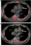
Better diagnostic certainty with DECT. A 38-year-old male with history of annual episodes of massive hemoptysis, which has been going on for several years and resolved each time with antibiotics and steroids and cough suppression. Initial axial conventional CT image (a) demonstrate a partially calcified mass within left aspect of the mediastinum (Arrow) at the expected location of left superior pulmonary vein (vein not visualized). The iodine density (b) and overlay images (c) demonstrate a large perfusion defect of the left upper lobe lung (Arrows), in the distribution of left superior pulmonary vein. Given the patent pulmonary arteries and above-mentioned findings, diagnosis of pulmonary venous occlusion secondary to granulomatous disease was considered.
Fig. 9
Fig. 9
Contrast boost by low keV VMI. A 32-year-old female, with a history of tricuspid atresia, status post left BT shunt in the newborn period, status post classic Fontan followed by fenestrated Fontan and subsequent left pulmonary artery reconstruction and placement of permanent pacemaker. The patient now presents with acute chest pain. Initial conventional CT image (a) demonstrate very poor opacification of the pulmonary arteries and a suspected filling defect/ embolus within the stented left main pulmonary artery. The low keV VM image (b) from the same exam provides the contrast boost and therefore diagnosis of sluggish flow within left pulmonary artery, secondary to the left superior pulmonary vein occlusion was made (Please note that left superior pulmonary vein is not visualized- consistent with chronic occlusion- Red arrow indicates expected location of left superior pulmonary vein and blue arrow represents a portion of left atrial appendage).
Fig. 10
Fig. 10
Contrast dose reduction with DECT. A 72-year-old male with history of renal dysfunction (eGFR =30 mL/min/1.73m2) comes for Pre – TAVI (Transcatheter Aortic valve Implantation) planning scan. (a) Conventional coronal CT image from DECT scan following intravenous administration of 25 mL of iodinated contrast shows poor contrast opacification of thoracic aorta. (b) Low keV VM image provides necessary contrast boost making the examination diagnostic at a very low contrast dose (as compared to 70-90 ml with conventional CT). Of note dense aortic valve calcification correlate with patient’s severe aortic stenosis (Arrow in a).
Fig. 11
Fig. 11
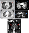
Improved lesion detection with DECT. A 79-year-old female with history of left upper lobe adenocarcinoma status post resection and radiation therapy returning for restaging scan. On conventional CT images in soft tissue and lung window settings (a, b), there is left apical post-surgical scarring seen without definite local recurrence. However, on iodine overlay image (c) there is focus of increased iodine accumulation within the scarring (Blue arrow), which correspond to increased radiotracer uptake on subsequent PET-CT fusion image (Red arrow in d). Pathology confirmed recurrent adenocarcinoma.
Fig. 12
Fig. 12
Better lesion characterization with DECT. A 74-year-old male with prior history of mediastinal mass consistent with small cell cancer, status post chemoradiotherapy. Initial conventional CT image (a) shows nonspecific fat stranding and soft tissue density in mediastinum, consistent with combination of tumor and post therapy changes. In addition, there is extensive collateralization in the mediastinum secondary to patients known Superior Vena Cava (SVC) involvement (Arrows in a). There is a new ill- defined hyperdense lesion seen in lower thoracic spine on conventional CT (Aarrow in b), which is indeterminate for metastatic disease. However, the vertebral lesion disappears on VNC image(c) pointing towards intraosseous enhancement secondary to SVC syndrome and not neoplastic lesion.
Fig. 13
Fig. 13
Incidental lesion assessment with DECT. A 80-year-old male was scanned as part of the pre TAVI (Transcatheter Aortic Valve Implantation) work up. Axial CT image (a) demonstrate incidentally noted hypodense (white arrows) and hyperdense left renal lesions(blue arrow), which were without iodine accumulation on iodine density (b)and Z effective images(c), consistent with hemorrhagic and non-hemorrhagic renal cysts.
Fig. 14
Fig. 14
Assessment of myocardial perfusion on DECT. A 81-year-old woman with history of chronic atrial fibrillation was planned for a left atrial appendage occlusion procedure. A planning cardiac CT; Early (a) and delayed conventional CT images (b) were unremarkable, however there was a clear large perfusion defect(Arrows), consistent with infarction, seen involving lateral left ventricular myocardium and papillary muscle on low keV VM image from delayed scan (c). Subsequent cardiac catheterization revealed total occlusion of the first obtuse marginal branch of the left circumflex coronary artery (not shown).
Fig. 15
Fig. 15
Radiation dose reduction with DECT. A 64-year-old male with history of endovascular aneurysm repair (EVAR) for abdominal aortic aneurysm (AAA), underwent a follow up three phase CT angiogram (non-contrast, arterial phase and delayed phase scanning) of the abdomen and pelvis. Axial arterial phase image (a) demonstrates areas of hyperattenuation outside the stent in the excluded abdominal aortic aneurysm (Arrows), which persists on true non-contrast (TNC) image(c) and does not change on delayed contrast enhanced image (b). The findings are consistent with calcification in the excluded AAA and not an endoleak. When utilizing the low keV VM image (d) and VNC (e) reconstructions from the single delayed phase examination (b), comparable information to a three-phase scan can be obtained, thus, there is a potential of significantly reducing radiation exposure to the patient.
Fig. 16
Fig. 16
Superior image quality with Photon-counting detector (PCD) CT. Coronal conventional CT (a) and PCD CT image (b) acquired in ultra-high resolution mode demonstrate better spatial resolution and delineation of coronary stent struts on PCD CT image, enabling reconstruction of a detailed three-dimensional volume rendered image(c). Images (Fig. 9a, 9b and 9c in original paper) reprinted with permission from S. Leng, M. Bruesewitz, S. Tao, K. Rajendran, A.F.Halaweish, N.G. Campeau, J.G. Fletcher, C.H. McCollough, Photon-counting Detector CT: System Design and Clinical Applications of an Emerging Technology. Radiographics. 39(3) (2019) 729–743.
None
None
None
None

References

    1. Takai M., Kaneko M. Discrimination between thorotrast and iodine contrast medium by means of dual-energy CT scanning. Phys. Med. Biol. 1984;29(8):959–967. - PubMed
    1. Kelcz F., Joseph P.M., Hilal S.K. Noise considerations in dual energy CT scanning. Med. Phys. 1979;6(5):418–425. - PubMed
    1. Yao Y., Ng J.M., Megibow A.J., Pelc N.J. Image quality comparison between single energy and dual energy CT protocols for hepatic imaging. Med. Phys. 2016;43(8):4877. - PubMed
    1. Heye T., Nelson R.C., Ho L.M., Marin D., Boll D.T. Dual-energy CT applications in the abdomen. AJR Am. J. Roentgenol. 2012;199(5 Suppl):S64–70. - PubMed
    1. Goo H.W., Goo J.M. Dual-energy C.T.: new horizon in medical imaging. Korean J. Radiol. 2017;18(4):555–569. - PMC - PubMed