. 2020 Sep 10;6(1):e10187.
doi: 10.1002/btm2.10187. eCollection 2021 Jan.

Cartilage-targeting ultrasmall lipid-polymer hybrid nanoparticles for the prevention of cartilage degradation

Affiliations

Cartilage-targeting ultrasmall lipid-polymer hybrid nanoparticles for the prevention of cartilage degradation

Xiangzhao Ai et al. Bioeng Transl Med. .

Abstract

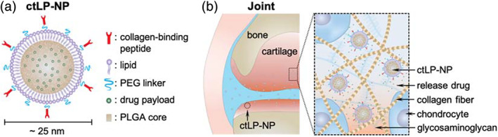
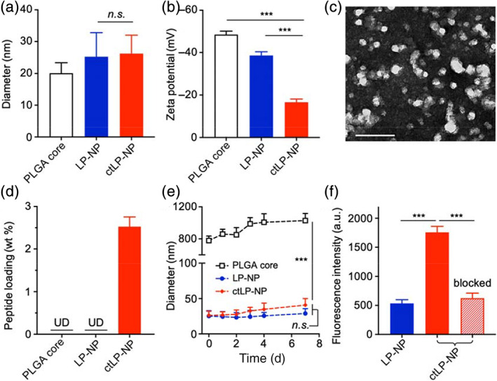
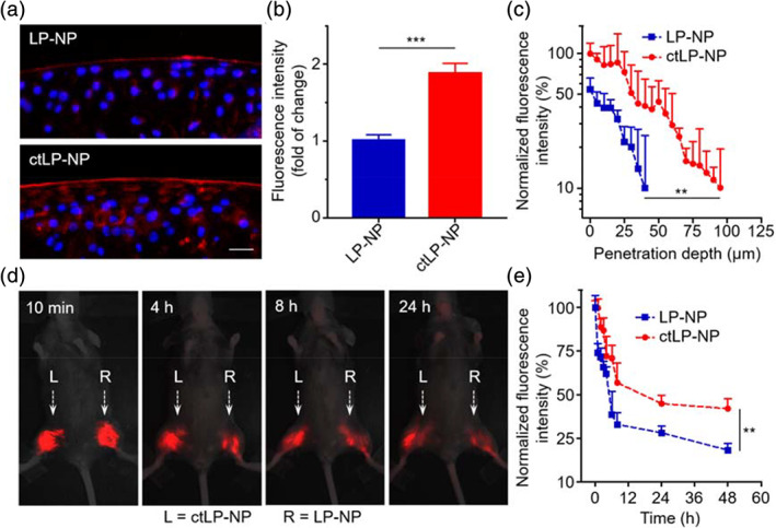
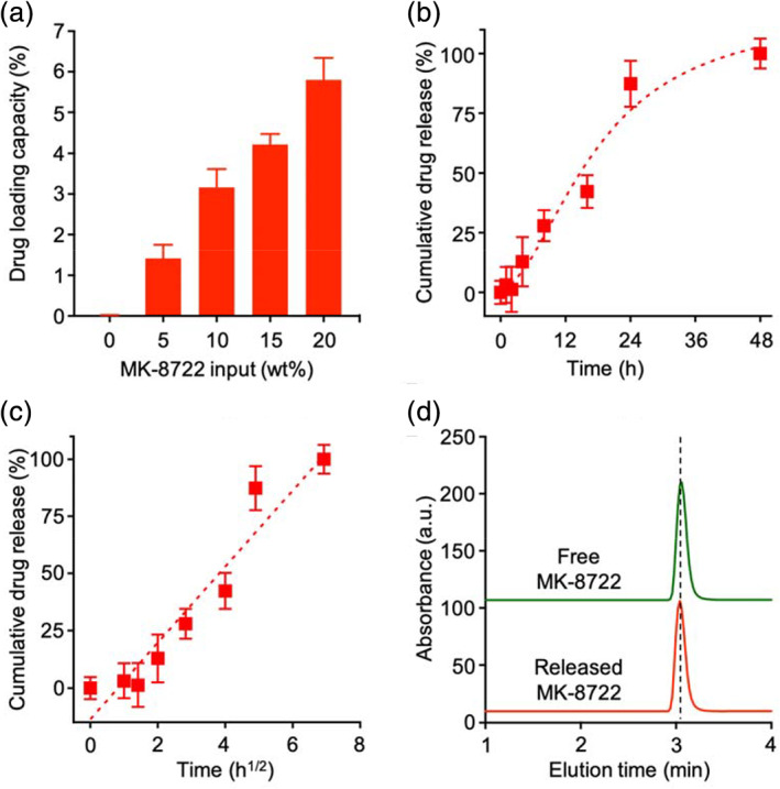
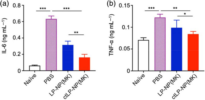
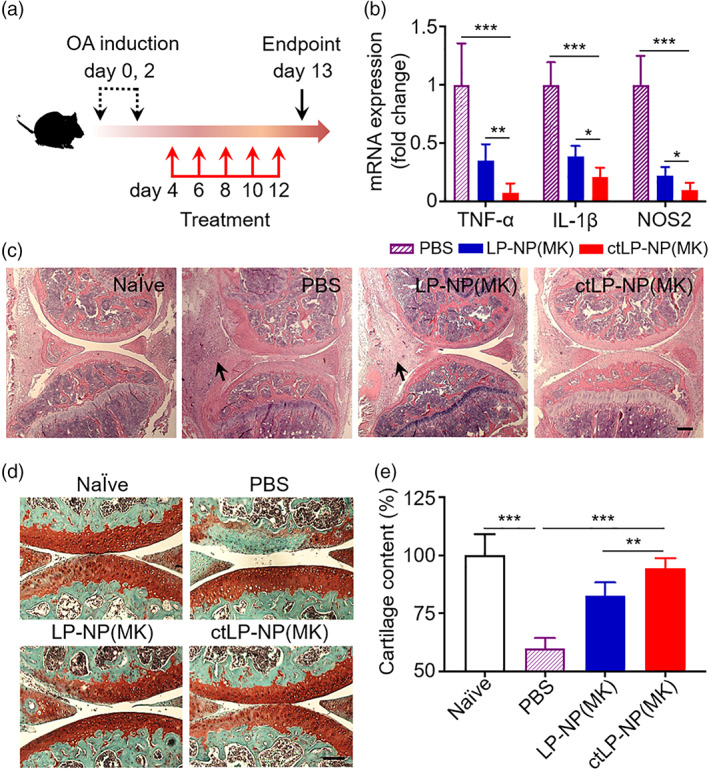
Current drug delivery approaches for the treatment of cartilage disorders such as osteoarthritis (OA) remain inadequate to achieve sufficient drug penetration and retention in the dense cartilage matrix. Herein, we synthesize sub-30 nm lipid-polymer hybrid nanoparticles functionalized with collagen-targeting peptides for targeted drug delivery to the cartilage. The nanoparticles consist of a polymeric core for drug encapsulation and a lipid shell modified with a collagen-binding peptide. By combining these design features, the nanoparticles can penetrate deep and accumulate preferentially in the cartilage. Using MK-8722, an activator of 5'-adenosine monophosphate-activated protein kinase (AMPK), as a model drug, the nanoparticles can encapsulate the drug molecules in high capacity and release them in a sustained and controllable manner. When injected into the knee joints of the mice with collagenase-induced OA, the drug-loaded nanoparticles can effectively reduce cartilage damage and alleviate the disease severity. Overall, the ultrasmall targeted nanoparticles represent a promising delivery platform to overcome barriers of dense tissues for the treatment of various indications, including cartilage disorders.

Keywords: intracartilage delivery; lipid‐polymer hybrid nanoparticle; nanomedicine; osteoarthritis; ultrasmall nanoparticle.


Conflict of interest statement

The authors declare no conflict of interest.

Figures

FIGURE 1
FIGURE 1
The design of collagen‐targeting ultrasmall lipid‐polymer hybrid nanoparticles (denoted “ctLP‐NPs”) for targeted drug delivery to the joints. (a) Schematic of ctLP‐NPs containing a core made from poly(lactic‐co‐glycolic acid) (PLGA) and a shell made from polyethylene glycol (PEG)‐conjugated lipid. For cartilage targeting, the nanoparticles are modified with collagen‐binding peptides. Hydrophobic drug molecules are encapsulated inside the PLGA cores for delivery. The nanoparticles have a small size around 25 nm. (b) The ctLP‐NPs are able to penetrate deep into the cartilage for effective drug targeting to the chondrocytes
FIGURE 2
FIGURE 2
Characterization of ctLP‐NPs. (a) Hydrodynamic size (diameter) of PLGA cores, LP‐NPs, and ctLP‐NPs, respectively. (b) Zeta potential (mV) of different nanoparticle groups. (c) A representative TEM image of ctLP‐NPs with uranyl acetate staining (scale bar, 50 nm). (d) Quantification of protein content on PLGA cores, LP‐NPs, and ctLP‐NPs, respectively, using a BCA assay (UD, undetectable). (e) The hydrodynamic size of PLGA cores, LP‐NPs, and ctLP‐NPs in 1× PBS over a week. (f) Fluorescence intensity of DiD‐labeled LP‐NPs and ctLP‐NPs bound onto a type II collagen‐coated plate. As an additional control (labeled with “blocked”), the plate was pretreated with free peptide to block the collagen before adding ctLP‐NPs. Data presented as mean ± SD (n = 3); n.s.: not significant; ***p < 0.001; statistical analysis by one‐way ANOVA
FIGURE 3
FIGURE 3
Retention and penetration of ctLP‐NPs in the cartilage. (a) Representative fluorescence images of femoral head sections incubated with DiD‐labeled LP‐NPs (top) or ctLP‐NPs (bottom). Red represents the nanoparticles and blue represents the nuclei (scale bars: 20 μm). (b) Relative fluorescence intensity in the cartilage upon incubation with LP‐NPs or ctLP‐NPs (0.2 mg mL−1) for 24 h. (c) Quantitative analysis of nanoparticle penetration depth into femoral heads. (d) Fluorescence images of mouse knee joints after intra‐articular injection of fluorescence‐labeled ctLP‐NPs (left, L) and LP‐NPs (right, R) at different timepoints. In the study, 20 μL of 2 mg mL−1 nanoparticle suspension was injected into each joint. (e) Fluorescence intensity of the nanoparticles in the knee joints. Data presented as mean ± SD (n = 3). **p < 0.01; ***p < 0.001; statistical analysis by two‐tailed Student's t‐test
FIGURE 4
FIGURE 4
Evaluation of ctLP‐NPs for MK‐8722 encapsulation and release. (a) The relationship between MK‐8722 initial input and its loading capacity. Data presented as mean + SD and n = 3. (b) A representative cumulative drug release profile of MK‐8722 from ctLP‐NPs in 1× PBS. (c) A plot of MK‐8722 release percentage from ctLP‐NPs against the square root of the release time. The linear fitting was made by using a diffusion‐dominant Higuchi model. (d) HPLC analysis of free MK‐8722 and released MK‐8722 from ctLP‐NPs, respectively. In (b) and (c), data presented as mean ± SD and n = 3
FIGURE 5
FIGURE 5
MK‐8722‐loaded ctLP‐NPs [ctLP‐NP(MK)] suppress inflammation in the cartilage. Mouse femoral heads were cultured with IL‐1β (10 ng mL−1) to induce inflammation. PBS, LP‐NP(MK), and ctLP‐NP(MK) were added to the tissue samples, respectively. After the incubation, levels of secreted cytokines including (a) IL‐6 and (b) TNF‐α were measured. Data presented as mean ± SD (n = 3); *p < 0.05, **p < 0.01, and ***p < 0.001; statistical analysis by one‐way ANOVA
FIGURE 6
FIGURE 6
Therapeutic efficacy of MK‐8722‐loaded ctLP‐NPs to repair cartilage damage in the collagenase‐induced OA (CIOA) mice. (a) The study protocol of therapeutic regimen within a CIOA mouse model. (b) Relative mRNA expression of cytokines (TNF‐α, IL‐1β, and NOS2) in the cartilage of mice knee joint upon PBS, LP‐NP(MK), and ctLP‐NP(MK) treatment (intra‐articular injection, 20 μL, 5 mg mL−1). (c) Representative images of H&E staining on knee joint sections from healthy mice (Naïve) and CIOA mice treated with PBS, LP‐NP(MK), and ctLP‐NP(MK), respectively. Scale bars, 100 μm. The arrows indicate areas where synovitis is discernible. (d) Representative images of safranin‐O staining on cartilage sections from healthy mice (Naïve) and CIOA mice treated with PBS LP‐NP(MK), and ctLP‐NP(MK), respectively. Scale bars, 100 μm. (e) Quantification of cartilage content from safranin‐O‐stained sections (red) in different groups. Data presented as mean ± SD (n = 3); **p < 0.01, ***p < 0.001; statistical analysis by one‐way ANOVA

References

    1. Litwic A, Edwards MH, Dennison EM, Cooper C. Epidemiology and burden of osteoarthritis. Br Med Bull. 2013;105:185‐199. - PMC - PubMed
    1. Vina ER, Kwoh CK. Epidemiology of osteoarthritis: literature update. Curr Opin Rheumatol. 2018;30:160‐167. - PMC - PubMed
    1. Allen KD, Golightly YM, White DK. Gaps in appropriate use of treatment strategies in osteoarthritis. Best Pract Res Clin Rheumatol. 2017;31:746‐759. - PubMed
    1. Loeser RF, Collins JA, Diekman BO. Ageing and the pathogenesis of osteoarthritis. Nat Rev Rheumatol. 2016;12:412‐420. - PMC - PubMed
    1. Mobasheri A, Rayman MP, Gualillo O, Sellam J, van der Kraan P, Fearon U. The role of metabolism in the pathogenesis of osteoarthritis. Nat Rev Rheumatol. 2017;13:302‐311. - PubMed