. 2021 Jan 7;24(2):102037.
doi: 10.1016/j.isci.2021.102037. eCollection 2021 Feb 19.

Impaired formation of high-order gephyrin oligomers underlies gephyrin dysfunction-associated pathologies

Affiliations

Impaired formation of high-order gephyrin oligomers underlies gephyrin dysfunction-associated pathologies

Seungjoon Kim et al. iScience. .

Abstract

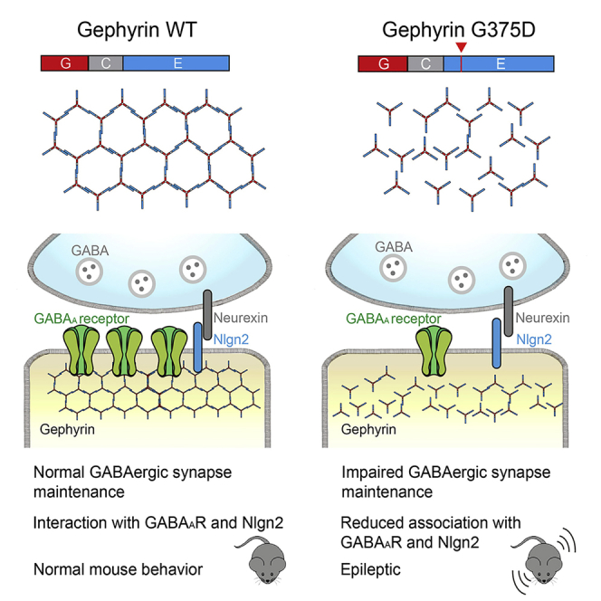
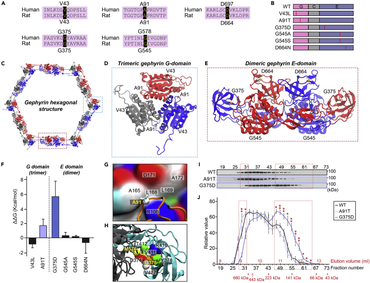
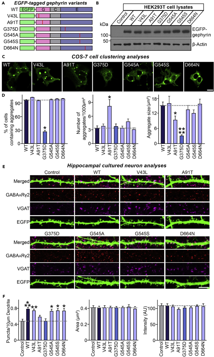
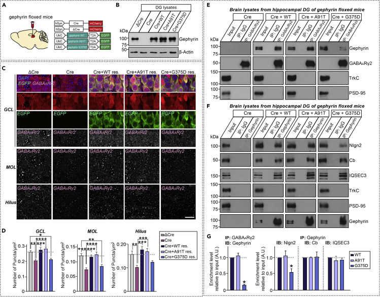
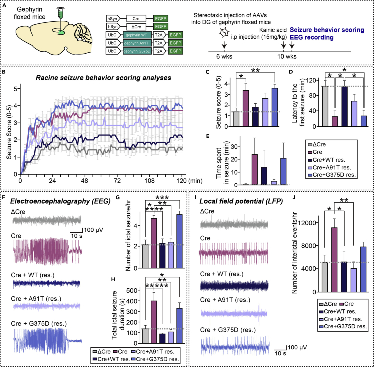
Gephyrin is critical for the structure, function, and plasticity of inhibitory synapses. Gephyrin mutations have been linked to various neurological disorders; however, systematic analyses of the functional consequences of these mutations are lacking. Here, we performed molecular dynamics simulations of gephyrin to predict how six reported point mutations might change the structural stability and/or function of gephyrin. Additional in silico analyses revealed that the A91T and G375D mutations reduce the binding free energy of gephyrin oligomer formation. Gephyrin A91T and G375D displayed altered clustering patterns in COS-7 cells and nullified the inhibitory synapse-promoting effect of gephyrin in cultured neurons. However, only the G375D mutation reduced gephyrin interaction with GABAA receptors and neuroligin-2 in mouse brain; it also failed to normalize deficits in GABAergic synapse maintenance and neuronal hyperactivity observed in hippocampal dentate gyrus-specific gephyrin-deficient mice. Our results provide insights into biochemical, cell-biological, and network-activity effects of the pathogenic G375D mutation.

Keywords: Molecular Biology; Neuroscience; Structural Biology.


Conflict of interest statement

The authors declare that they have no conflicts of interest.

Figures

None
Graphical abstract
Figure 1
Figure 1
A human gephyrin missense mutation associated with epileptic encephalitis alters multimer binding stability (A) Alignment and conservation between human gephyrin- and rat gephyrin residues that are mutated in human patients with epileptic encephalopathy or ASDs. (B) Schematic diagrams of gephyrin WT and its mutants. Abbreviations: G, G-domain; C, C-domain; E, E-domain. (C) Schematic depiction of gephyrin hexagonal lattices. (D) Structure of G-domain trimers and position of point mutations in gephyrin. (E) Structure of E-domain dimers and position of point mutations in gephyrin. (F) Multimer binding free energy difference between gephyrin WT and the indicated point mutants, as calculated by thermodynamic integration analyses. Numerical values per mutation points were averaged. Data are represented as means ± SDs. (G) A91 and hydrophobic pockets in the binding interface between gephyrin G-domains. (H) G375 positioned inside of the β-strand bundle at the binding interface between chains. Bead and surface colors indicate the following: white, hydrophobic; green, hydrophilic; red, negatively charged; blue, positively charged; and yellow, methionine. (I) Extracts of HEK293T cells transfected with untagged gephyrin WT or its mutants (A91T or G375D) were fractionated on a gel filtration column. Fractions were analyzed by immunoblotting using anti-gephyrin antibodies. (J) Quantification of expression levels of gephyrin WT and its mutants (A91T or G375D) in each fraction. Data are means ± SEMs (∗p < 0.05, ∗∗p < 0.01; WT versus G375D; Mann-Whitney U test; n = 5/group).
Figure 2
Figure 2
Gephyrin G375D, but not A91T, is defective in promoting GABAergic synapse formation in cultured hippocampal neurons (A) Schematic diagrams of gephyrin WT and its mutants. Abbreviations: G, G-domain; C, C-domain; E, E-domain. (B) Immunoblotting of lysates from HEK293T cells transfected with EGFP-tagged gephyrin WT or its mutants using anti-gephyrin antibodies. An anti-β-actin antibody was used as a normalization control. (C) Representative images of COS-7 cells transfected with EGFP-gephyrin WT or its mutants. Scale bar, 10 μm (applies to all images). (D) Quantification of the percentage of COS-7 cells with large cytoplasmic intracellular aggregates, the number of aggregates per 1000 μm2, and average aggregate size. Data are presented as means ± SEMs from three independent experiments (∗p < 0.05, ∗∗∗∗p < 0.0001; non-parametric ANOVA with Kruskal-Wallis test followed by post hoc Dunn's multiple comparison test). (E) Representative images of cultured hippocampal neurons transfected at DIV10 with EGFP alone (Control) or cotransfected with EGFP alone and EGFP-tagged gephyrin WT or its mutants, and analyzed at DIV14 by triple-immunofluorescence staining using anti-GABAARγ2 (red), anti-VGAT (magenta) and anti-EGFP (green) antibodies. Scale bar, 10 μm (applies to all images). (F) Summary graphs of the effects of overexpressing gephyrin WT or its mutants on GABAARγ2+VGAT+ puncta density (left), GABAARγ2+VGAT+ puncta size (middle), and GABAARγ2+VGAT+ puncta intensity (right). Data are presented as means ± SEMs from three independent experiments (2–3 dendrites per transfected neurons were analyzed and group-averaged; ∗p < 0.05, ∗∗p < 0.01, ∗∗∗p < 0.001; non-parametric ANOVA with Kruskal-Wallis test followed by post hoc Dunn's multiple comparison test).
Figure 3
Figure 3
Gephyrin G375D impairs promotion of GABAergic synapse maintenance in vivo (A) Schematic diagram of AAV vectors expressing Cre or ΔCre and WT gephyrin and its mutants (A91T and G375D) used for stereotactic injection into the DG of gephyrin floxed mice. (B) Immunoblotting analyses with gephyrin antibodies validating gephyrin knockout and expression of gephyrin rescue constructs in vivo. Lysates from mouse brains stereotactically injected with AAVs were collected and immunoblotted with anti-gephyrin antibodies. An anti-β-actin antibody was used as a normalization control. (C) Representative images showing GABAARγ2+ puncta in the DG of mice stereotactically injected with the indicated AAVs. Scale bar, 20 μm (applies to all images). Abbreviations: MOL, molecular layer; GCL, granule cell layer. (D) Quantification of the density of GABAARγ2+ puncta per tissue area. Data are presented as means ± SEMs (n = 4 mice each after averaging data from 5 sections/mouse; ∗p < 0.05, ∗∗p < 0.01, ∗∗∗p < 0.001, ∗∗∗∗p < 0.0001; non-parametric ANOVA with Kruskal-Wallis test followed by post hoc Dunn's multiple comparison test). (E) Synaptosomal fractions of adult brains from gephyrin floxed mice stereotactically injected with the indicated AAV viruses were immunoprecipitated with anti-GABAARγ2 antibodies and immunoblotted with anti-GABAARγ2, anti-gephyrin, anti-TrkC or anti-PSD-95 antibodies. An equal amount of rabbit IgG was used as a negative control. Input, 5%. (F) Synaptosomal fractions of adult brains from gephyrin floxed mice stereotactically injected with the indicated AAV viruses were immunoprecipitated with an anti-gephyrin antibody and immunoblotted with the indicated antibodies. An equal amount of mouse IgG was used as a negative control. Input, 5%. (G) Quantification of coimmunoprecipitated synaptic proteins in (E) or (F), normalized to controls. Data are means ± SEM from three independent experiments. (∗p < 0.05; non-parametric Kruskal-Wallis test with Dunn's post hoc test).
Figure 4
Figure 4
Gephyrin G375D mutant fails to rescue increased seizure susceptibility in DG-specific gephyrin-cKO mice (A) Schematic diagram of AAV vectors expressing Cre or ΔCre and WT gephyrin or its mutants (A91T and G375D) used for stereotactic injection into the DG of gephyrin floxed mice. Experimental scheme for seizure scoring and EEG recordings. The DG region of the hippocampus of ~6-week-old gephyrin floxed mice was bilaterally injected with AAVs-ΔCre or Cre, or co-injected with Cre- and gephyrin-WT–expressing AAVs (Cre + WT res.) or gephyrin-mutant–expressing AAVs (Cre + A91T res. or Cre + G375D res.). Mice were intraperitoneally administered KA 2 weeks after AAV injections, after which seizures were scored and EEGs were recorded. (B) KA-induced seizures in mice injected with the indicated AAVs were scored every 3 min for a total of 120 min, as described in Supplemental Information. Data are presented as means ± SEMs (ΔCre, n = 7 mice; Cre, n = 7 mice; Cre + WT [res.], n = 7 mice; Cre + A91T [res.], n = 7 mice; and Cre + G375D [res.], n = 7 mice; ∗p < 0.05, ∗∗p < 0.01, ∗∗∗p < 0.001 vs. control; Kruskal-Wallis test followed by Dunn's post hoc test). (C) Quantification of mean score values under each experimental condition. Data are presented as means ± SEMs (n = 7 mice/condition; ∗p < 0.05, ∗∗p < 0.01 vs. control; Kruskal-Wallis test followed by Dunn's post hoc test). (D) Quantification of latency to the first seizure after KA administration under each condition. Data are presented as means ± SEMs (n = 7 mice/condition; ∗p < 0.05; Kruskal-Wallis test followed by Dunn's post hoc test). (E) Quantification of time spent in seizure under each condition. Data are presented as means ± SEMs (n = 7 mice/condition). (F) Representative EEG traces of ictal-like seizures recorded from the cortex under the indicated experimental conditions. (G and H) Quantification of the number of ictal-like seizures (G) and total duration of ictal-like seizures (H) per hour under each condition. Data are presented as means ± SEMs (n = 8–11 mice/condition; ∗p < 0.05, ∗∗p < 0.01, ∗∗∗p < 0.001 vs. control; Kruskal-Wallis test followed by Dunn's post hoc test). (I) Representative LFP traces of inter-ictal events recorded from the DG under the indicated experimental conditions. (J) Quantification of the number of inter-ictal events under each condition. Data are presented as means ± SEMs (n = 5–6 mice/condition; ∗p < 0.05, ∗∗p < 0.01 vs. control; Kruskal-Wallis test followed by Dunn's post hoc test).

Similar articles

Cited by

References

    1. Agarwal S., Tannenberg R.K., Dodd P.R. Reduced expression of the inhibitory synapse scaffolding protein gephyrin in Alzheimer's disease. J. Alzheimers Dis. 2008;14:313–321. - PubMed
    1. Alvarez F.J. Gephyrin and the regulation of synaptic strength and dynamics at glycinergic inhibitory synapses. Brain Res. Bull. 2017;129:50–65. - PubMed
    1. Anastasia A., Deinhardt K., Chao M.V., Will N.E., Irmady K., Lee F.S., Hempstead B.L., Bracken C. Val66Met polymorphism of BDNF alters prodomain structure to induce neuronal growth cone retraction. Nat. Commun. 2013;4:2490. - PMC - PubMed
    1. Antonelli R., Pizzarelli R., Pedroni A., Fritschy J.M., Del Sal G., Cherubini E., Zacchi P. Pin1-dependent signalling negatively affects GABAergic transmission by modulating neuroligin2/gephyrin interaction. Nat. Commun. 2014;5:5066. - PMC - PubMed
    1. Bath K.G., Lee F.S. Variant BDNF (Val66Met) impact on brain structure and function. Cogn. Affect. Behav. Neurosci. 2006;6:79–85. - PubMed

LinkOut - more resources