[Preprint]. 2021 Jan 27:2021.01.27.428466.
doi: 10.1101/2021.01.27.428466.

Synthetic nanobody-SARS-CoV-2 receptor-binding domain structures identify distinct epitopes

Affiliations

Synthetic nanobody-SARS-CoV-2 receptor-binding domain structures identify distinct epitopes

Javeed Ahmad et al. bioRxiv. .

Update in

Abstract

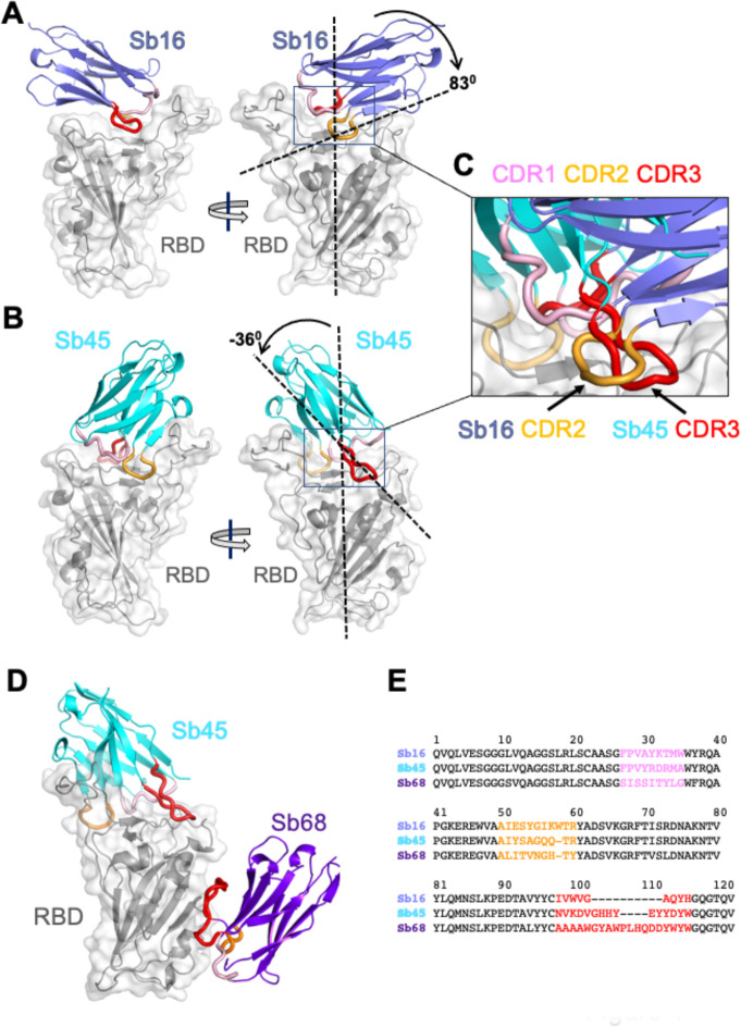
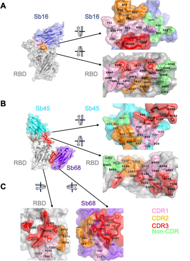
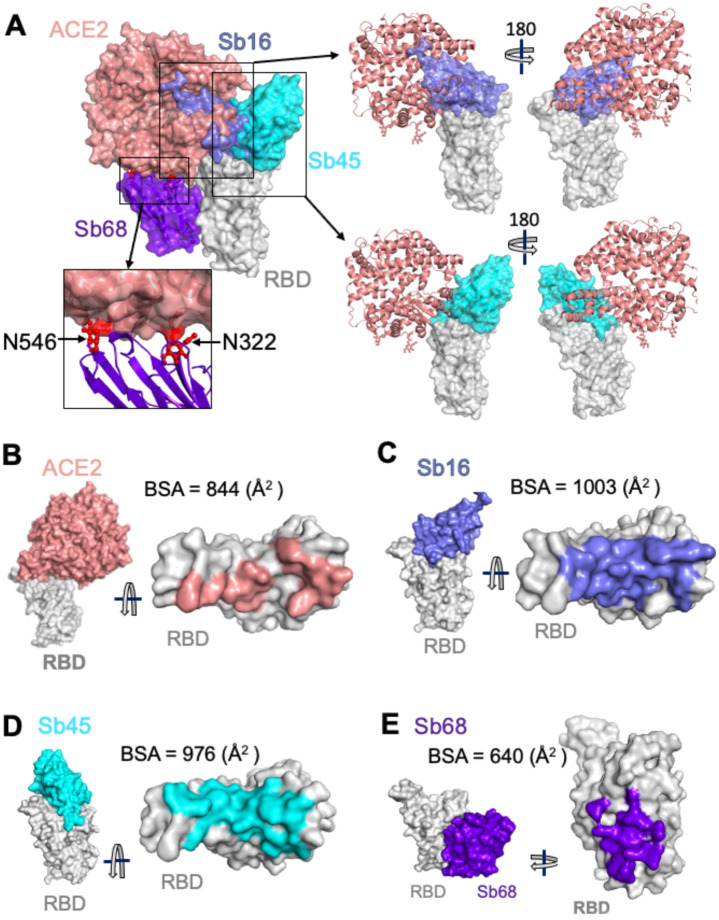
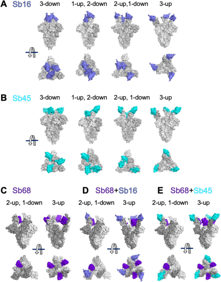
The worldwide spread of severe acute respiratory syndrome coronavirus 2 (SARS-CoV-2) demands unprecedented attention. We report four X-ray crystal structures of three synthetic nanobodies (sybodies) (Sb16, Sb45 and Sb68) bind to the receptor-binding domain (RBD) of SARS-CoV-2: binary complexes of Sb16-RBD and Sb45-RBD; a ternary complex of Sb45-RBD-Sb68; and Sb16 unliganded. Sb16 and Sb45 bind the RBD at the ACE2 interface, positioning their CDR2 and CDR3 loops diametrically. Sb16 reveals a large CDR2 shift when binding the RBD. Sb68 interacts peripherally at the ACE2 interface; steric clashes with glycans explain its mechanism of viral neutralization. Superposing these structures onto trimeric spike (S) protein models indicates these sybodies bind conformations of the mature S protein differently, which may aid therapeutic design.

One sentence summary: X-ray structures of synthetic nanobodies complexed with the receptor-binding domain of the spike protein of SARS-CoV-2 reveal details of CDR loop interactions in recognition of distinct epitopic sites.


Conflict of interest statement

Competing interests: The authors declare no competing interests.

Figures

Fig. 1.
Fig. 1.
Overall structures of Sb16, Sb45 and Sb68 complexes with SARS-CoV-2 RBD. Ribbons (sybodies) and ribbons plus surface (RBD) representations of the complex of (A) Sb16 (slate) with RBD (grey) (7KGK); (B) Sb45 (cyan) with RBD (7KGJ), and (C) Sb45 and Sb68 (purple) with RBD (7KLW). Sb16-RBD and Sb45-RBD, superimposed based on the RBD are shown in (D) to highlight CDR loops, which are color coded as indicated. The CDR2 of Sb16 and CDR3 of Sb45 interact similarly with the RBD surface.
Fig. 2.
Fig. 2.
Interface and interaction of (A) Sb16-RBD, (B) Sb45-RBD and (C) Sb68-RBD. (Individual contacting residues are listed in TableS2 in Supplemental Materials). CDR1, CDR2, CDR3 regions are painted pink, orange and red respectively. Additional non-CDR region contacting residues are colored lime. On the RBD surface, the epitopic residues that contact the sybodies are colored according to the sybody CDR.
Fig. 3.
Fig. 3.
Sybodies compete with ACE2 for RBD binding. (A) Sb16 (slate), Sb45 (cyan) and Sb68 (purple) - RBD complexes were superposed on the ACE2—RBD structure (salmon) (6M0J) based on the RBD. Sb16 is buried inside ACE2; Sb45 is partially buried in ACE2; and Sb68 has major clashes with two N-glycan sites (N322 and N546) of ACE2. (B) Epitopic areas (on RBD) captured by ACE2 (salmon), BSA = 844 (Å2); (C) by Sb16 (slate), BSA = 1003 (Å2); (D) by Sb45 (cyan), BSA = 976 (Å2); and (E) by Sb68 (purple), BSA = 640 (Å2).
Fig. 4.
Fig. 4.
Superposition of complexes on spike models reveals accessibility of sybodies. Superposition of (A) Sb16-RBD (slate) on spike (6XEY: 3-down; 6Z43: 1-up and 2-down; 7A29: 2-up and 1-down; 7JVC: 3-up); (B) Sb45-RBD (cyan); and (C) Sb68-RBD (purple), there is no accessible surface for Sb68 on 3-down of spike; (D) Sb68 and Sb16 on RBD; (E) SB68 and Sb45 on RBD.

References

    1. Conti P. et al., The British variant of the new coronavirus-19 (Sars-Cov-2) should not create a vaccine problem. J Biol Regul Homeost Agents. 2021. (10.23812/21-3-E). - DOI - PubMed
    1. Kirby T., New variant of SARS-CoV-2 in UK causes surge of COVID-19. Lancet Respir Med. 2021. (10.1016/S2213-2600(21)00005-9). - DOI - PMC - PubMed
    1. Tang J. W., Tambyah P. A., Hui D. S., Emergence of a new SARS-CoV-2 variant in the UK. J Infect. 2020. (10.1016/j.jinf.2020.12.024). - DOI - PMC - PubMed
    1. Wibmer C. K. et al., SARS-CoV-2 501Y.V2 escapes neutralization by South African COVID-19 donor plasma. bioRxiv, 2021.2001.2018.427166 (2021). - PubMed
    1. Wang Q. et al., Structural and Functional Basis of SARS-CoV-2 Entry by Using Human ACE2. Cell. 2020. (10.1016/j.cell.2020.03.045). - DOI - PMC - PubMed

Publication types