[Preprint]. 2021 Feb 12:2021.01.25.428137.
doi: 10.1101/2021.01.25.428137.

Antibody Resistance of SARS-CoV-2 Variants B.1.351 and B.1.1.7

Affiliations

Antibody Resistance of SARS-CoV-2 Variants B.1.351 and B.1.1.7

Pengfei Wang et al. bioRxiv. .

Update in

  • Antibody resistance of SARS-CoV-2 variants B.1.351 and B.1.1.7.
    Wang P, Nair MS, Liu L, Iketani S, Luo Y, Guo Y, Wang M, Yu J, Zhang B, Kwong PD, Graham BS, Mascola JR, Chang JY, Yin MT, Sobieszczyk M, Kyratsous CA, Shapiro L, Sheng Z, Huang Y, Ho DD. Wang P, et al. Nature. 2021 May;593(7857):130-135. doi: 10.1038/s41586-021-03398-2. Epub 2021 Mar 8. Nature. 2021. PMID: 33684923

Abstract

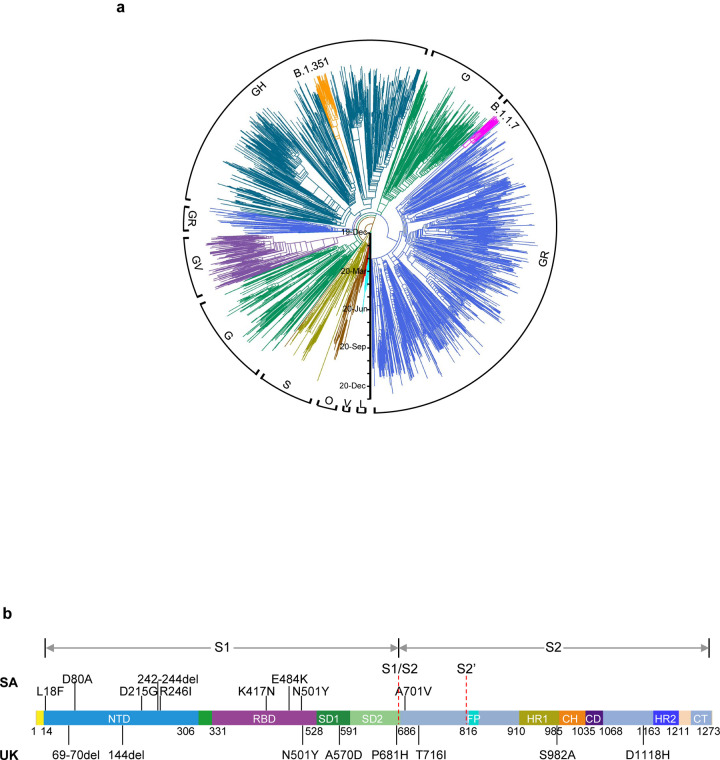
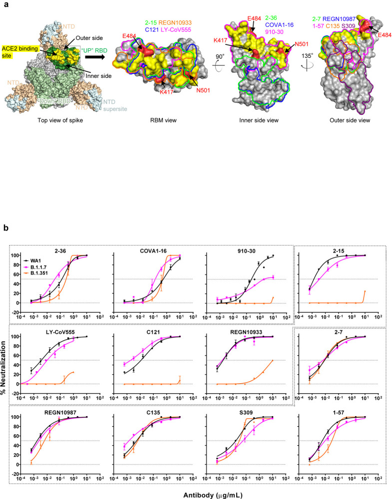
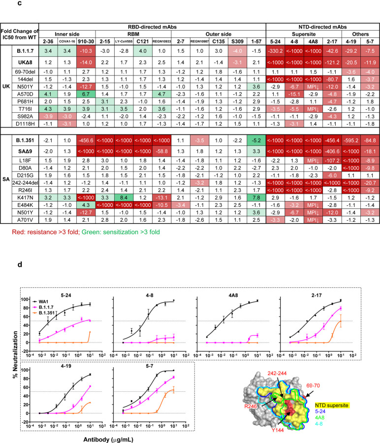
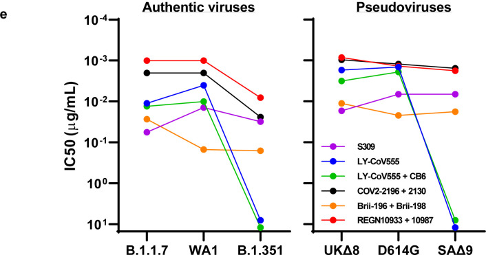
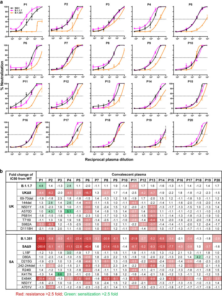
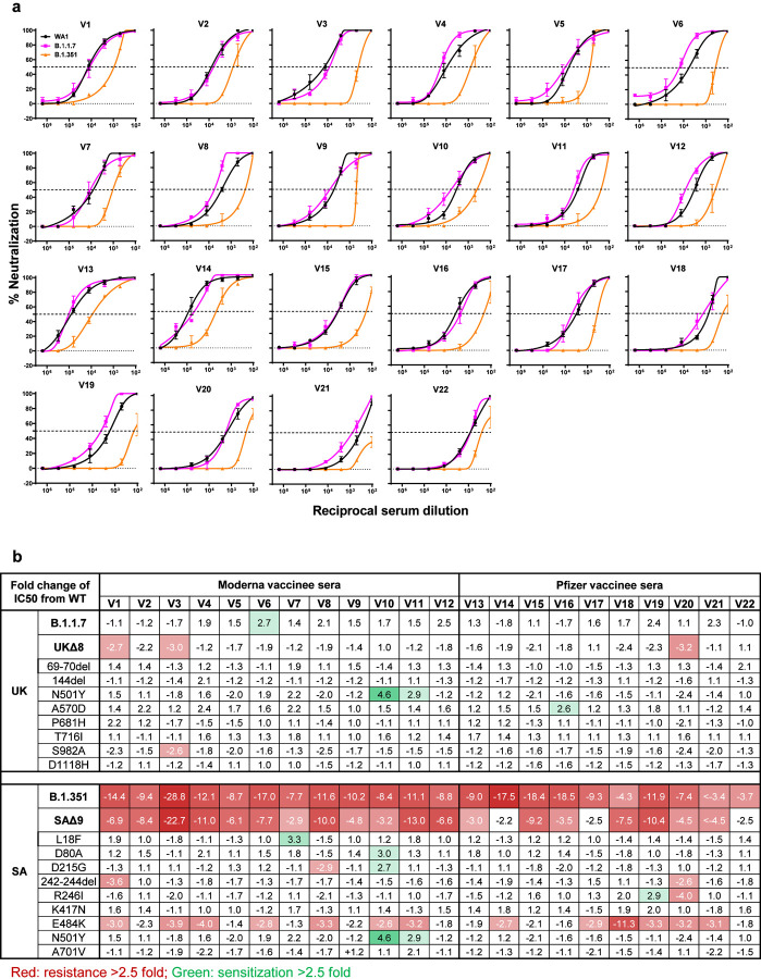
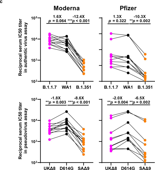
The COVID-19 pandemic has ravaged the globe, and its causative agent, SARS-CoV-2, continues to rage. Prospects of ending this pandemic rest on the development of effective interventions. Single and combination monoclonal antibody (mAb) therapeutics have received emergency use authorization1-3, with more in the pipeline4-7. Furthermore, multiple vaccine constructs have shown promise8, including two with ~95% protective efficacy against COVID-199,10. However, these interventions were directed toward the initial SARS-CoV-2 that emerged in 2019. The recent emergence of new SARS-CoV-2 variants B.1.1.7 in the UK11 and B.1.351 in South Africa12 is of concern because of their purported ease of transmission and extensive mutations in the spike protein. We now report that B.1.1.7 is refractory to neutralization by most mAbs to the N-terminal domain (NTD) of spike and relatively resistant to a few mAbs to the receptor-binding domain (RBD). It is not more resistant to convalescent plasma or vaccinee sera. Findings on B.1.351 are more worrisome in that this variant is not only refractory to neutralization by most NTD mAbs but also by multiple individual mAbs to the receptor-binding motif on RBD, largely due to an E484K mutation. Moreover, B.1.351 is markedly more resistant to neutralization by convalescent plasma (9.4 fold) and vaccinee sera (10.3-12.4 fold). B.1.351 and emergent variants13,14 with similar spike mutations present new challenges for mAb therapy and threaten the protective efficacy of current vaccines.


Figures

Fig. 1 |
Fig. 1 |. Emerging SARS-CoV-2 variants identified in the United Kingdom and South Africa.
a, Phylogenetic tree of SARS-CoV-2 variants, with B.1.351 and B.1.1.7 highlighted. b, Mutations in the viral spike identified in B.1.351 (SA) and B.1.1.7 (UK) in addition to D614G.
Fig. 2 |
Fig. 2 |. Susceptibility of B.1.1.7 and B.1.351 to neutralization by mAbs.
a, Footprints of neutralizing mAbs on the RBD. Left panel, top view of SARS-COV-2 spike with one RBD in the “up” conformation (pdb: 6zgg). RBD and NTD are colored green and peach, respectively. The positions of ‘inner’ and ‘outer’ sides are indicated on the “up” RBD with the ACE2-binding site colored yellow. The three panels to the right show the antibody footprints on RBD. b, Neutralization of B.1.1.7, B.1.351, and WT viruses by select RBD mAbs. c, Fold increase or decrease in IC50 of neutralizing mAbs against B.1.1.7 and B.1.351, as well as UKΔ8, SAΔ9, and single-mutation pseudoviruses, relative to WT, presented as a heatmap with darker colors implying greater change. MPI↓ denotes that maximum percent inhibition is substantially reduced, confounding IC50 calculations. d, Neutralization of B.1.1.7, B.1.351, and WT viruses by NTD-directed mAbs, the footprints of which are delineated by the color tracings in the insert. e, Changes in neutralization IC50 of authorized or investigational therapeutic mAbs against B.1.1.7, B.1.351, WT (WA1) viruses as well as UKΔ8, SAΔ9, and WT (D614G) pseudoviruses. Data in b and d are mean ± SEM of technical triplicates, and represent one of two independent experiments.
Fig. 3 |
Fig. 3 |. B.1.351 is more resistant to neutralization by convalescent plasma from patients.
a, Neutralization results for 20 convalescent plasma samples (P1–P20) against B.1.1.7, B.1.351, and WT viruses. Data represent mean ± SEM of technical triplicates. b, Fold increase or decrease in neutralization IC50 of B.1.1.7 and B.1.351, as well as UKΔ8, SAΔ9, and single-mutation pseudoviruses, relative to the WT presented as a heatmap with darker colors implying greater change. c, Change in reciprocal plasma neutralization IC50 values of convalescent plasma against B.1.1.7 and B.1.351, as well as UKΔ8 and SAΔ9, relative to the WT. Mean fold changes in IC50 values relative to the WT are written above the p values. Statistical analysis was performed using a Wilcoxon matched-pairs signed rank test. Two-tailed p-values are reported.
Fig. 4 |
Fig. 4 |. B.1.351 is more resistant to neutralization by vaccinee sera.
a, Neutralization profiles for 22 serum samples obtained from persons who received SARS-CoV-2 vaccine made by Moderna (V1-V12) or Pfizer (V13-V22) against B.1.1.7, B.1.351, and WT viruses. Data are mean ± SEM of technical triplicates, and represent one of two independent experiments. b, Fold change in serum neutralization IC50 of B.1.1.7 and B.1.351, as well as UKΔ8, SAΔ9, and single-mutation pseudoviruses, relative to the WT, presented as a heatmap with darker colors implying greater change. c, Change in reciprocal serum IC50 values for Moderna and Pfizer vaccinees against B.1.1.7 and B.1.351, as well as UKΔ8 and SAΔ9, relative to the WT. Mean fold change in IC50 relative to the WT is written above the p values. Statistical analysis was performed using a Wilcoxon matched-pairs signed rank test. Two-tailed p-values are reported.

References

    1. Chen P. et al. SARS-CoV-2 Neutralizing Antibody LY-CoV555 in Outpatients with Covid-19. N Engl J Med 384, 229–237 (2021). - PMC - PubMed
    1. Hansen J. et al. Studies in humanized mice and convalescent humans yield a SARS-CoV-2 antibody cocktail. Science 369, 1010–1014 (2020). - PMC - PubMed
    1. Gottlieb R. L. et al. Effect of Bamlanivimab as Monotherapy or in Combination With Etesevimab on Viral Load in Patients With Mild to Moderate COVID-19: A Randomized Clinical Trial. JAMA (2021). - PMC - PubMed
    1. Ju B. et al. Human neutralizing antibodies elicited by SARS-CoV-2 infection. Nature 584, 115–119 (2020). - PubMed
    1. Pinto D. et al. Cross-neutralization of SARS-CoV-2 by a human monoclonal SARS-CoV antibody. Nature 583, 290–295 (2020). - PubMed

Publication types