

Genome-wide analysis identifies novel susceptibility loci for myocardial infarction
- PMID: 33532862
- PMCID: PMC7936531
- DOI: 10.1093/eurheartj/ehaa1040
Genome-wide analysis identifies novel susceptibility loci for myocardial infarction
Abstract
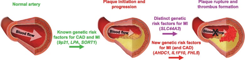
Aims: While most patients with myocardial infarction (MI) have underlying coronary atherosclerosis, not all patients with coronary artery disease (CAD) develop MI. We sought to address the hypothesis that some of the genetic factors which establish atherosclerosis may be distinct from those that predispose to vulnerable plaques and thrombus formation.
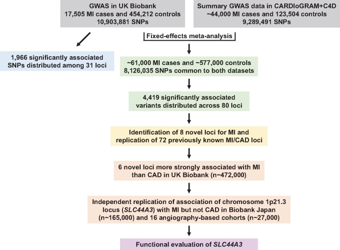
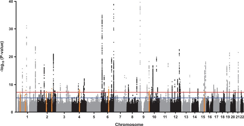
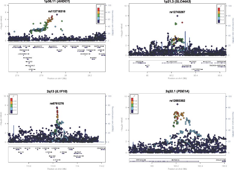
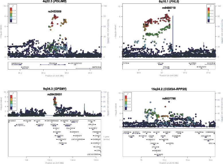
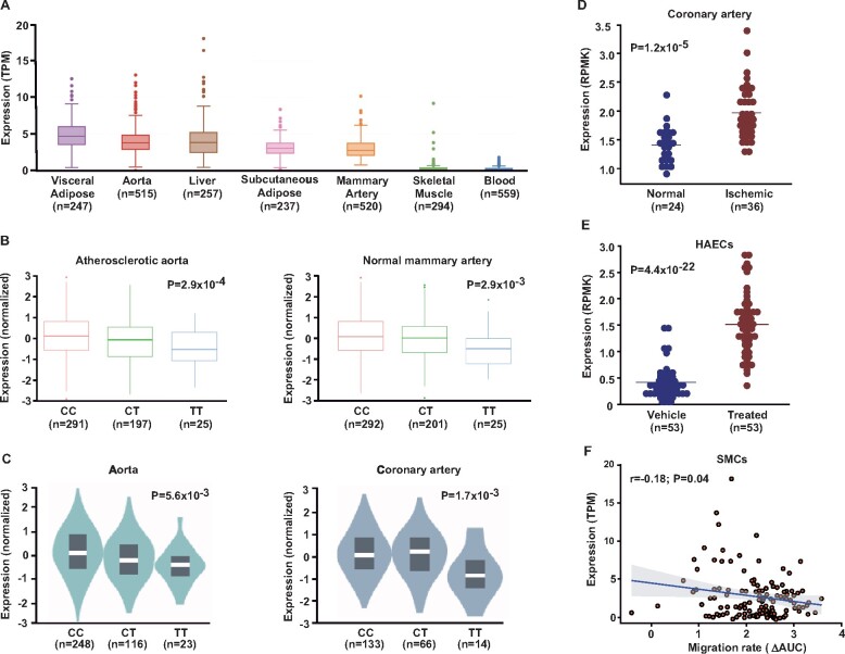
Methods and results: We carried out a genome-wide association study for MI in the UK Biobank (n∼472 000), followed by a meta-analysis with summary statistics from the CARDIoGRAMplusC4D Consortium (n∼167 000). Multiple independent replication analyses and functional approaches were used to prioritize loci and evaluate positional candidate genes. Eight novel regions were identified for MI at the genome wide significance level, of which effect sizes at six loci were more robust for MI than for CAD without the presence of MI. Confirmatory evidence for association of a locus on chromosome 1p21.3 harbouring choline-like transporter 3 (SLC44A3) with MI in the context of CAD, but not with coronary atherosclerosis itself, was obtained in Biobank Japan (n∼165 000) and 16 independent angiography-based cohorts (n∼27 000). Follow-up analyses did not reveal association of the SLC44A3 locus with CAD risk factors, biomarkers of coagulation, other thrombotic diseases, or plasma levels of a broad array of metabolites, including choline, trimethylamine N-oxide, and betaine. However, aortic expression of SLC44A3 was increased in carriers of the MI risk allele at chromosome 1p21.3, increased in ischaemic (vs. non-diseased) coronary arteries, up-regulated in human aortic endothelial cells treated with interleukin-1β (vs. vehicle), and associated with smooth muscle cell migration in vitro.
Conclusions: A large-scale analysis comprising ∼831 000 subjects revealed novel genetic determinants of MI and implicated SLC44A3 in the pathophysiology of vulnerable plaques.
Keywords: Genetic factors; Genome-wide association study; Meta-analysis; Myocardial infarction; SLC44A3.
Published on behalf of the European Society of Cardiology. All rights reserved. © The Author(s) 2021. For permissions, please email: journals.permissions@oup.com.
Figures
Comment in
-
Unfolding and disentangling coronary vascular disease through genome-wide association studies.Eur Heart J. 2021 Mar 1;42(9):934-937. doi: 10.1093/eurheartj/ehaa1089. Eur Heart J. 2021. PMID: 33561196 Free PMC article. No abstract available.
References
-
- Go AS, Mozaffarian D, Roger VL, Benjamin EJ, Berry JD, Blaha MJ, Dai S, Ford ES, Fox CS, Franco S, Fullerton HJ, Gillespie C, Hailpern SM, Heit JA, Howard VJ, Huffman MD, Judd SE, Kissela BM, Kittner SJ, Lackland DT, Lichtman JH, Lisabeth LD, Mackey RH, Magid DJ, Marcus GM, Marelli A, Matchar DB, McGuire DK, Mohler ER 3rd, Moy CS, Mussolino ME, Neumar RW, Nichol G, Pandey DK, Paynter NP, Reeves MJ, Sorlie PD, Stein J, Towfighi A, Turan TN, Virani SS, Wong ND, Woo D, Turner MB; American Heart Association Statistics Committee and Stroke Statistics Subcommittee. Heart disease and stroke statistics—2014 update: a report from the American Heart Association. Circulation 2014;129:e28–e292. - PMC - PubMed
-
- Ridker PM, Danielson E, Fonseca FA, Genest J, Gotto AM Jr, Kastelein JJ, Koenig W, Libby P, Lorenzatti AJ, MacFadyen JG, Nordestgaard BG, Shepherd J, Willerson JT, Glynn RJ; JUPITER Study Group. Rosuvastatin to prevent vascular events in men and women with elevated C-reactive protein. N Engl J Med 2008;359:2195–2207. - PubMed
-
- Hansson GK. Inflammation, atherosclerosis, and coronary artery disease. N Engl J Med 2005;352:1685–1695. - PubMed
-
- Schunkert H, Erdmann J, Samani NJ. Genetics of myocardial infarction: a progress report. Eur Heart J 2010;31:918–925. - PubMed
-
- Erdmann J, Stark K, Esslinger UB, Rumpf PM, Koesling D, de Wit C, Kaiser FJ, Braunholz D, Medack A, Fischer M, Zimmermann ME, Tennstedt S, Graf E, Eck S, Aherrahrou Z, Nahrstaedt J, Willenborg C, Bruse P, Brænne I, Nöthen MM, Hofmann P, Braund PS, Mergia E, Reinhard W, Burgdorf C, Schreiber S, Balmforth AJ, Hall AS, Bertram L, Steinhagen-Thiessen E, Li S-C, März W, Reilly M, Kathiresan S, McPherson R, Walter UCARDIoGRAMOtt J, Samani NJ, Strom TM, Meitinger T, Hengstenberg C, Schunkert H. Dysfunctional nitric oxide signalling increases risk of myocardial infarction. Nature 2013;504:432–436. - PubMed
Publication types
MeSH terms
Grants and funding
- R01 ES021801/ES/NIEHS NIH HHS/United States
- R01 HL125863/HL/NHLBI NIH HHS/United States
- R01 HL128300/HL/NHLBI NIH HHS/United States
- R01 HL126827/HL/NHLBI NIH HHS/United States
- R01 HL113147/HL/NHLBI NIH HHS/United States
- P01 HL076491/HL/NHLBI NIH HHS/United States
- R01 HL142856/HL/NHLBI NIH HHS/United States
- R01 HL148239/HL/NHLBI NIH HHS/United States
- K24 HL107643/HL/NHLBI NIH HHS/United States
- R21 HL135230/HL/NHLBI NIH HHS/United States
- FS/14/76/30933/BHF_/British Heart Foundation/United Kingdom
- R01 HL103931/HL/NHLBI NIH HHS/United States
- R01 HL147187/HL/NHLBI NIH HHS/United States
- R01 HL150359/HL/NHLBI NIH HHS/United States
- R01 MD010358/MD/NIMHD NIH HHS/United States
- R01 HL144651/HL/NHLBI NIH HHS/United States
- P30 ES006694/ES/NIEHS NIH HHS/United States
- R00 HL138193/HL/NHLBI NIH HHS/United States
- P01 HL147823/HL/NHLBI NIH HHS/United States
- P01 ES022845/ES/NIEHS NIH HHS/United States
- R01 HL103866/HL/NHLBI NIH HHS/United States
- R01 HL135920/HL/NHLBI NIH HHS/United States
- R01 HL133169/HL/NHLBI NIH HHS/United States
- P01 HL098055/HL/NHLBI NIH HHS/United States
- R01 HL147883/HL/NHLBI NIH HHS/United States
- R01 HL148110/HL/NHLBI NIH HHS/United States
- R01 ES025786/ES/NIEHS NIH HHS/United States
LinkOut - more resources
Full Text Sources
Other Literature Sources
Medical
Miscellaneous
