

Proposal of a scoring system for predicting pathological risk based on a semiautomated analysis of whole slide images in oral squamous cell carcinoma
- PMID: 33533145
- PMCID: PMC8247849
- DOI: 10.1002/hed.26621
Proposal of a scoring system for predicting pathological risk based on a semiautomated analysis of whole slide images in oral squamous cell carcinoma
Abstract
Background: The study aimed to evaluate the risk factors based on pathological findings comprehensively in oral squamous cell carcinoma (OSCC) using image analysis.
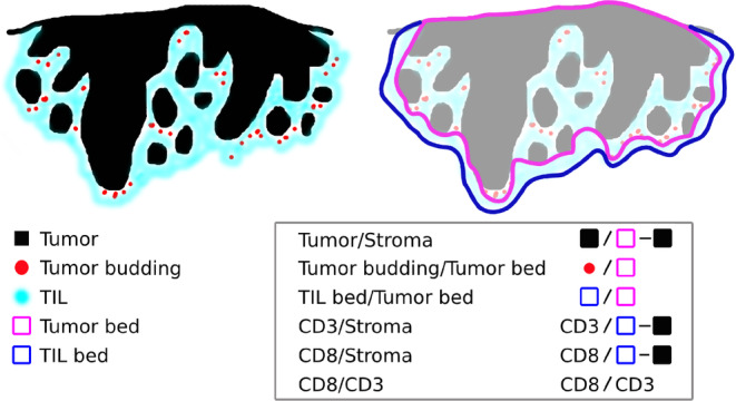
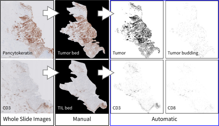
Methods: Scanned images of hematoxylin and eosin-, pan-cytokeratin-, CD3-, and CD8-stained slides of OSCC cases from 256 patients were analyzed, and six variables were obtained including the tumor-stroma ratio, tumor budding per tumor bed area, and tumor infiltrating lymphocytes-associated variables. We determined the "score" of all cases based on the variables, and all cases were classified into low-, intermediate-, and high-risk groups.
Results: A significant difference in prognosis was confirmed between the risk groups (p < 0.001), and even when evaluated within different tumor-node-metastasis (TNM) stages, the high-risk groups were associated with poor survival.
Conclusions: We report our work on a possible descriptive model that can predict prognosis based on pathological and imaging findings regardless of the TNM stage.
Keywords: image analysis; oral squamous cell carcinoma; tumor budding; tumor infiltrating lymphocytes; whole slide image.
© 2021 The Authors. Head & Neck published by Wiley Periodicals LLC.
Conflict of interest statement
The author declares that there is no conflict of interest that could be perceived as prejudicing the impartiality of the research reported.
Figures
Similar articles
-
Exploring the combination of tumor-stroma ratio, tumor-infiltrating lymphocytes, and tumor budding with WHO histopathological grading on early-stage oral squamous cell carcinoma prognosis.J Oral Pathol Med. 2023 May;52(5):402-409. doi: 10.1111/jop.13359. Epub 2022 Oct 3. J Oral Pathol Med. 2023. PMID: 36149755
-
Novel pathological predictive factors for extranodal extension in oral squamous cell carcinoma: a retrospective cohort study based on tumor budding, desmoplastic reaction, tumor-infiltrating lymphocytes, and depth of invasion.BMC Cancer. 2022 Apr 13;22(1):402. doi: 10.1186/s12885-022-09393-8. BMC Cancer. 2022. PMID: 35418058 Free PMC article.
-
Prognostic significance of tumor infiltrating immune cells in oral squamous cell carcinoma.BMC Cancer. 2017 May 26;17(1):375. doi: 10.1186/s12885-017-3317-2. BMC Cancer. 2017. PMID: 28549420 Free PMC article.
-
Association of Stathmin (Op18) with TNM Staging and Grading of Oral Squamous Cell Carcinoma and Its Role in Tumor Progression.J Contemp Dent Pract. 2022 May 1;23(5):497-502. J Contemp Dent Pract. 2022. PMID: 35986456
-
Tumor-infiltrating ICOS+ Effector Regulatory T-Cells in Oral Squamous Cell Carcinoma as a Promising Biomarker for Prognosis and 'Hot' Tumor.Anticancer Res. 2022 May;42(5):2383-2393. doi: 10.21873/anticanres.15717. Anticancer Res. 2022. PMID: 35489733 Review.
Cited by
-
Predictive value of tumor budding in head and neck squamous cell carcinoma: an update.Virchows Arch. 2023 Oct;483(4):441-449. doi: 10.1007/s00428-023-03630-6. Epub 2023 Aug 29. Virchows Arch. 2023. PMID: 37642731 Review.
-
The Emerging Impact of Tumor Budding in Oral Squamous Cell Carcinoma: Main Issues and Clinical Relevance of a New Prognostic Marker.Cancers (Basel). 2022 Jul 22;14(15):3571. doi: 10.3390/cancers14153571. Cancers (Basel). 2022. PMID: 35892830 Free PMC article. Review.
-
Prognostic Significance of Tumor-Stroma Ratio (TSR) in Head and Neck Squamous Cell Carcinoma: Systematic Review and Meta-Analysis.Cells. 2024 Oct 26;13(21):1772. doi: 10.3390/cells13211772. Cells. 2024. PMID: 39513879 Free PMC article.
-
Artificial Intelligence for Image Analysis in Oral Squamous Cell Carcinoma: A Review.Diagnostics (Basel). 2023 Jul 20;13(14):2416. doi: 10.3390/diagnostics13142416. Diagnostics (Basel). 2023. PMID: 37510160 Free PMC article. Review.
-
Motivation for Smoking Cessation in Patients With Oral Squamous Cell Carcinoma-A One-Time Survey.Clin Exp Dent Res. 2025 Aug;11(4):e70154. doi: 10.1002/cre2.70154. Clin Exp Dent Res. 2025. PMID: 40788291 Free PMC article.
References
-
- Ferlay J, Colombet M, Soerjomataram I, et al. Estimating the global cancer incidence and mortality in 2018: GLOBOCAN sources and methods. Int J Cancer. 2019;144:1941‐1953. - PubMed
-
- American Joint Committee on Cancer . Cancer Staging Manual. 8th ed. New York: Springer; 2017.
-
- Vangangelt KMH, Tollenaar LSA, van Pelt GW, et al. The prognostic value of tumor–stroma ratio in tumor‐positive axillary lymph nodes of breast cancer patients. Int J Cancer. 2018;143:3194‐3200. - PubMed
-
- Aurello P, Berardi G, Giulitti D, et al. Tumor–stroma ratio is an independent predictor for overall survival and disease free survival in gastric cancer patients. Surgeon. 2017;15:329‐335. - PubMed
MeSH terms
LinkOut - more resources
Full Text Sources
Other Literature Sources
Medical
Research Materials
