. 2021 Mar;11(3):753-767.
doi: 10.1002/2211-5463.13107. Epub 2021 Feb 18.

Environmental pH stress influences cellular secretion and uptake of extracellular vesicles

Affiliations

Environmental pH stress influences cellular secretion and uptake of extracellular vesicles

Ikuhiko Nakase et al. FEBS Open Bio. 2021 Mar.

Abstract

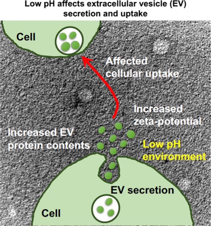
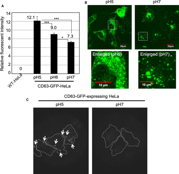
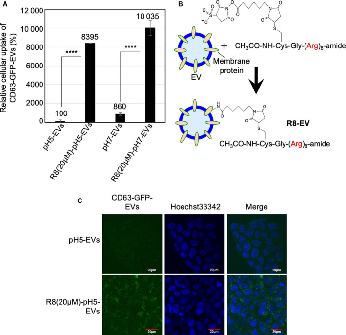
Exosomes (extracellular vesicles/EVs) participate in cell-cell communication and contain bioactive molecules, such as microRNAs. However, the detailed characteristics of secreted EVs produced by cells grown under low pH conditions are still unknown. Here, we report that low pH in the cell culture medium significantly affected the secretion of EVs with increased protein content and zeta potential. The intracellular expression level and location of stably expressed GFP-fused CD63 (an EV tetraspanin) in HeLa cells were also significantly affected by environmental pH. In addition, increased cellular uptake of EVs was observed. Moreover, the uptake rate was influenced by the presence of serum in the cell culture medium. Our findings contribute to our understanding of the effect of environmental conditions on EV-based cell-cell communication.

Keywords: cell-penetrating peptides; cellular uptake; exosomes; extracellular vesicles; pH.


Conflict of interest statement

The authors declare no conflict of interest.

Figures

Fig. 1
Fig. 1
Schematic of our research findings: low pH affects EV secretion and cellular uptake.
Fig. 2
Fig. 2
Effects of low pH cell culture condition on expression of CD63‐GFP fusion proteins. (A) Relative fluorescent intensity of CD63‐GFP fusion proteins stably expressing in HeLa cells cultured for 24 h at 37 °C in α‐MEM (pH 7, 6 or 5) with 10% FBS, analyzed using a flow cytometer (detection for 10 000 live cells). The data are expressed as the mean (± SD) of three experiments. Differences between groups were compared by one‐way ANOVA with Tukey's post hoc test. *P < 0.05, ***P < 0.001. (B) Confocal microscopic observation of CD63‐GFP fusion proteins stably expressing HeLa cells cultured for 24 h at 37 °C in α‐MEM (pH 7 or 5) with 10% FBS. Scale bar, 20 μm (enlarged pictures: 10 μm). (C) TIRF observation of CD63‐GFP fusion proteins stably expressing HeLa cells cultured for 24 h at 37 °C in α‐MEM (pH 7 or 5) with 10% FBS. Arrows show comparatively high fluorescent intensity of CD63‐GFP fusion protein expression in comparison with that at pH 7 cell culture condition. Dotted white lines show the cellular structures.
Fig. 3
Fig. 3
Increased protein content of secreted EVs under low pH cell culture condition. (A) Relative protein concentration of EVs/cell number under different pH conditions (pH 5, 6, or 7) cell culture condition. EVs secreted from CD63‐GFP‐HeLa cells (48 h, 37 °C) were analyzed by BCA protein assay, and each protein concentration was divided by the cell number. The data are expressed as the mean (± SD) of three experiments. Differences between groups were compared by one‐way ANOVA with Dunnett's post hoc test. *P < 0.05. (B) Particle images and number of secreted EVs were detected by NanoSight. (C) Average diameter and zeta potential of the isolated CD63‐GFP‐EVs secreted from CD63‐GFP‐HeLa under same cell culture condition as that in (A). (D) TEM observation of CD63‐GFP‐EVs secreted by CD63‐GFP‐HeLa cells under pH 5 condition of (A). Scale bar: 500 nm (left) and 200 nm (right, enlarged).
Fig. 4
Fig. 4
Increased cellular uptake (serum‐starved condition) of isolated EVs secreted under low pH cell culture condition. (A) Confocal microscopic observation of A431 cells treated with isolated CD63‐GFP‐EVs [20 μg·mL−1 for 48 h at 37 °C in MEM (pH 7) without FBS], secreted under pH 7 or pH 5 cell culture condition. Blue signals, Hoechst 33342 for nuclear staining; green signals, CD63‐GFP‐EVs. Scale bar, 20 μm. (B) The relative cellular uptake of isolated CD63‐GFP‐EVs [20 μg·mL−1 for 48 h at 37 °C in MEM (pH 7 or 5) without FBS] secreted under pH 7 or pH 5 cell culture condition, analyzed using a flow cytometer (detection for 10 000 live cells). The data are expressed as the mean (± SD) of three experiments. Differences between groups were compared by two‐way ANOVA followed by Bonferroni's post hoc test ***P < 0.001, ****P < 0.0001.
Fig. 5
Fig. 5
Effect of serum and modification of octaarginine (R8) peptides on cellular EV uptake. (A) The relative cellular uptake (A431 cells) of isolated CD63‐GFP‐EVs [20 μg·mL−1 for 24 h at 37 °C in MEM (pH 7) with 10% FBS] secreted under pH 7 or pH 5 cell culture condition, analyzed using a flow cytometer (detection for 10 000 live cells). The data are expressed as the mean (± SD) of three experiments. Differences between groups were compared by two‐way ANOVA followed by Bonferroni's post hoc test. ****P < 0.0001. (B) Modification of R8 peptides on exosomal membrane via suffo‐EMCS linker. (C) Confocal microscopic observation of A431 cells treated with isolated CD63‐GFP‐EVs [20 μg·mL−1 for 24 h at 37 °C in MEM (pH 7) with FBS] secreted under pH 5 cell culture condition, with or without R8 peptide modification on EV membrane. Blue signals, Hoechst 33342 for nuclear staining; green signals, CD63‐GFP‐EVs. Scale bar, 20 μm.

References

    1. Tan A, Rajadas J and Seifalian AM (2013) Exosomes as nano‐theranostic delivery platforms for gene therapy. Adv Drug Deliv Rev 65, 357–367. - PubMed
    1. Tai YL, Chen KC, Hsieh JT and Shen TL (2018) Exosomes in cancer development and clinical applications. Cancer Sci 109, 2364–2374. - PMC - PubMed
    1. Hannafon BN and Ding WQ (2013) Intercellular communication by exosome‐derived microRNAs in cancer. Int J Mol Sci 14, 14240–14269. - PMC - PubMed
    1. Escudier B, Dorval T, Chaput N, André F, Caby MP, Novault S, Flament C, Leboulaire C, Borg C, Amigorena S et al. (2005) Vaccination of metastatic melanoma patients with autologous dendritic cell (DC) derived‐exosomes: results of the first phase I clinical trial. J Transl Med 3, 10. - PMC - PubMed
    1. Morse MA, Garst J, Osada T, Khan S, Hobeika A, Clay TM, Valente N, Shreeniwas R, Sutton MA, Delcayre A et al. (2005) A phase I study of dexosome immunotherapy in patients with advanced non‐small cell lung cancer. J Transl Med 3, 9. - PMC - PubMed

Publication types