Early events in light chain aggregation at physiological pH reveal new insights on assembly, stability, and aggregate dissociation
- PMID: 33533277
- PMCID: PMC8848840
- DOI: 10.1080/13506129.2021.1877129
Early events in light chain aggregation at physiological pH reveal new insights on assembly, stability, and aggregate dissociation
Abstract
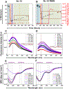
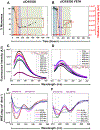
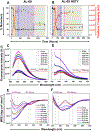
Early events in immunoglobulin light chain (AL) amyloid formation are especially important as some early intermediates formed during the aggregation reaction are cytotoxic and play a critical role in the initiation of amyloid assembly. We investigated the early events in in vitro aggregation of cardiac amyloidosis AL proteins at pH 7.4. In this study we make distinctions between general aggregation and amyloid formation. Aggregation is defined by the disappearance of monomers and the detection of sedimentable intermediates we call non-fibrillar macromolecular (NFM) intermediates by transmission electron microscopy (TEM). Amyloid formation is defined by the disappearance of monomers, Thioflavin T fluorescence enhancement, and the presence of fibrils by TEM. All proteins aggregated at very similar rates via the formation of NFM intermediates. The condensed NFM intermediates were composed of non-native monomers. Amyloid formation and amyloid yield was variable among the different proteins. During the stationary phase, all proteins demonstrated different degrees of dissociation. These dissociated species could play a key role in the already complex pathophysiology of AL amyloidosis. The degree of dissociation is inversely proportional to the amyloid yield. Our results highlight the importance and physiological consequences of intermediates/fibril dissociation in AL amyloidosis.
Keywords: Immunoglobulin light chain; Thioflavin T; aggregate dissociation; circular dichroism; intrinsic fluorescence; light chain amyloidosis; non-fibrillar macromolecular intermediates.
Conflict of interest statement
Disclosure of interest:
The authors declare that they have no conflicts of interest with the contents of this article.
Figures


Similar articles
-
Mechanistic Insights into the Early Events in the Aggregation of Immunoglobulin Light Chains.Biochemistry. 2019 Jul 23;58(29):3155-3168. doi: 10.1021/acs.biochem.9b00311. Epub 2019 Jul 9. Biochemistry. 2019. PMID: 31287666 Free PMC article.
-
Partially folded intermediates as critical precursors of light chain amyloid fibrils and amorphous aggregates.Biochemistry. 2001 Mar 27;40(12):3525-35. doi: 10.1021/bi001782b. Biochemistry. 2001. PMID: 11297418
-
Aggregation of Full-length Immunoglobulin Light Chains from Systemic Light Chain Amyloidosis (AL) Patients Is Remodeled by Epigallocatechin-3-gallate.J Biol Chem. 2017 Feb 10;292(6):2328-2344. doi: 10.1074/jbc.M116.750323. Epub 2016 Dec 28. J Biol Chem. 2017. PMID: 28031465 Free PMC article.
-
The Ultrastructure of Tissue Damage by Amyloid Fibrils.Molecules. 2021 Jul 29;26(15):4611. doi: 10.3390/molecules26154611. Molecules. 2021. PMID: 34361762 Free PMC article. Review.
-
Immunoglobulin light chain amyloid aggregation.Chem Commun (Camb). 2018 Sep 20;54(76):10664-10674. doi: 10.1039/c8cc04396e. Chem Commun (Camb). 2018. PMID: 30087961 Free PMC article. Review.
Cited by
-
Mechanistic insights into the aggregation pathway of the patient-derived immunoglobulin light chain variable domain protein FOR005.Nat Commun. 2023 Jun 23;14(1):3755. doi: 10.1038/s41467-023-39280-0. Nat Commun. 2023. PMID: 37353525 Free PMC article.
-
Biophysical characterization of human-cell-expressed, full-length κI O18/O8, AL-09, λ6a, and Wil immunoglobulin light chains.Biochim Biophys Acta Proteins Proteom. 2024 May 1;1872(3):140993. doi: 10.1016/j.bbapap.2023.140993. Epub 2023 Dec 31. Biochim Biophys Acta Proteins Proteom. 2024. PMID: 38169170 Free PMC article.
-
A bibliometric analysis of light chain amyloidosis from 2005 to 2024: research trends and hot spots.Front Med (Lausanne). 2024 Jul 30;11:1441032. doi: 10.3389/fmed.2024.1441032. eCollection 2024. Front Med (Lausanne). 2024. PMID: 39139790 Free PMC article.
-
Role of the mechanisms for antibody repertoire diversification in monoclonal light chain deposition disorders: when a friend becomes foe.Front Immunol. 2023 Jul 13;14:1203425. doi: 10.3389/fimmu.2023.1203425. eCollection 2023. Front Immunol. 2023. PMID: 37520549 Free PMC article. Review.
-
Kinetic evidence for multiple aggregation pathways in antibody light chain variable domains.Protein Sci. 2024 Mar;33(3):e4871. doi: 10.1002/pro.4871. Protein Sci. 2024. PMID: 38100259 Free PMC article.
References
-
- Buxbaum J Mechanisms of disease: monoclonal immunoglobulin deposition. Amyloidosis, light chain deposition disease, and light and heavy chain deposition disease. Hematol Oncol Clin North Am. 1992. Apr;6(2):323–46. - PubMed
-
- Solomon A Light chains of human immunoglobulins. Methods Enzymol. 1985;116:101–21. - PubMed
MeSH terms
Substances
Grants and funding
LinkOut - more resources
Full Text Sources
Other Literature Sources
Medical
Miscellaneous
