. 2021 Apr;239(4):1073-1083.
doi: 10.1007/s00221-020-06030-3. Epub 2021 Feb 3.

The role of delta and theta oscillations during ego-motion in healthy adult volunteers

Affiliations

The role of delta and theta oscillations during ego-motion in healthy adult volunteers

M Ertl et al. Exp Brain Res. 2021 Apr.

Abstract

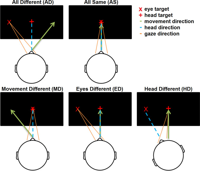
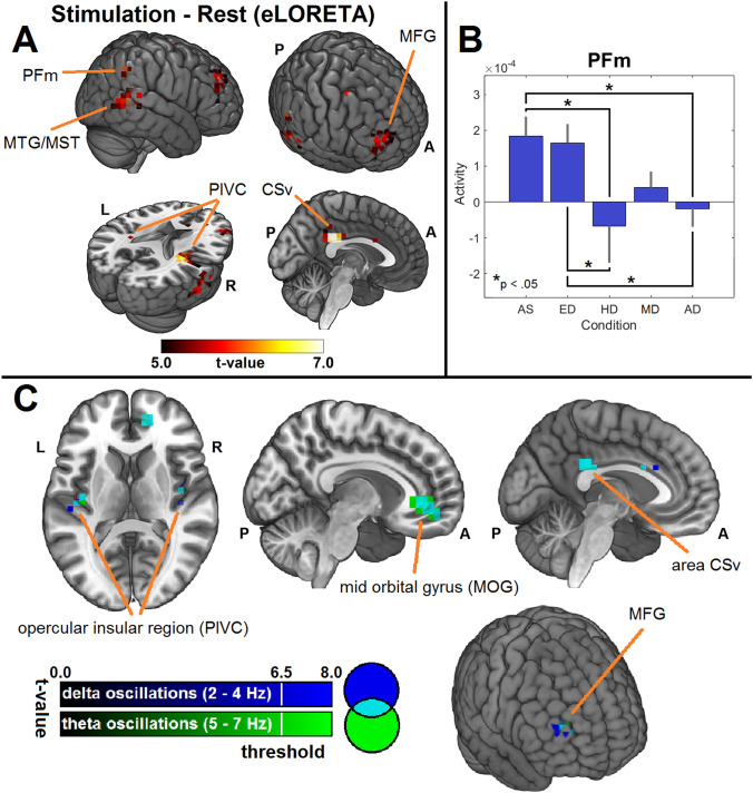
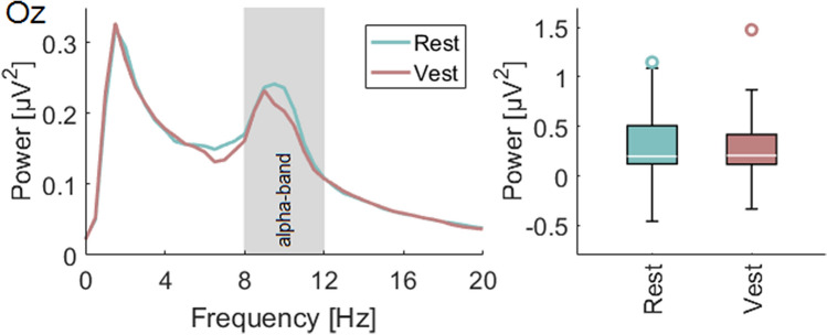
The successful cortical processing of multisensory input typically requires the integration of data represented in different reference systems to perform many fundamental tasks, such as bipedal locomotion. Animal studies have provided insights into the integration processes performed by the neocortex and have identified region specific tuning curves for different reference frames during ego-motion. Yet, there remains almost no data on this topic in humans.In this study, an experiment originally performed in animal research with the aim to identify brain regions modulated by the position of the head and eyes relative to a translational ego-motion was adapted for humans. Subjects sitting on a motion platform were accelerated along a translational pathway with either eyes and head aligned or a 20° yaw-plane offset relative to the motion direction while EEG was recorded.Using a distributed source localization approach, it was found that activity in area PFm, a part of Brodmann area 40, was modulated by the congruency of translational motion direction, eye, and head position. In addition, an asymmetry between the hemispheres in the opercular-insular region was observed during the cortical processing of the vestibular input. A frequency specific analysis revealed that low-frequency oscillations in the delta- and theta-band are modulated by vestibular stimulation. Source-localization estimated that the observed low-frequency oscillations are generated by vestibular core-regions, such as the parieto-opercular region and frontal areas like the mid-orbital gyrus and the medial frontal gyrus.

Keywords: Alpha activity; Hemispheric asymmetry; Multisensory; Passive motion; Reference frames; Vestibular stimulation; Vestibular system.


Conflict of interest statement

The authors declare that they have no conflict of interest.

Figures

Fig. 1
Fig. 1
Visualization of the head, and eye position relative to the platform movement during the five conditions. In the “all different” (AD) condition the head-, eye, and movement all pointed into three different directions with an angle of 20° between them. During the “all same” (AS) condition the eyes, head, and movement directions were all aligned. During the other three conditions either the movement (MD), eyes (ED), head (HD) positions deviated from the remaining two. The positions of the targets varied between trials and all possible combinations were presented during the experiment
Fig. 2
Fig. 2
a Acceleration profile used for this experiment. b Grand Average of all conditions and subjects. Due to different vestibular thresholds across the subjects and due to a less restrictive head fixation the VestEPs described by other studies (P1, N1, P2) are less pronounced. c Time–frequency representation of the time course. A strong signal increase during the movement can be observed in the delta (1–4 Hz) and theta-band (4–7 Hz)
Fig. 3
Fig. 3
a Results of the source localization analysis. We found a network consisting of the opercular-insular cortex (PIVC), area CSv, the middle temporal gyrus (MTG), the medial frontal gyrus (MFG) and area PFm, a sub-region of Brodmann area 40, as main generators during the movement compared to the rest period. Comparing the activity in left and right PIVC a hemispheric asymmetry with stronger activity in the right hemisphere can be seen. b Only the activity in area PFm was significantly modulated by the relative positions of the eyes, head, and movement. The activity was particularly decreased in conditions in which the head was not aligned with the eyes and movement. c Frequency-specific source localization analysis for frequencies in the delta- and theta-band comparing the activity during motion to the rest period. The results revealed activity in the cortical vestibular “core network” (PIVC, area CSv) and frontal structures (MOG, MFG)
Fig. 4
Fig. 4
Fourier-Transformation of the signal recorded at electrode Oz for frequencies between 0 and 20 Hz during the rest period and the vestibular stimulation (Vest) elicited by the platform movement. Comparing the alpha power for the individual alpha-frequencies of the subjects a slight but not significant decrease can be observed during motion compared to rest

Similar articles

Cited by

References

    1. Andersen RA, Snyder LH, Bradley DC, Xing J. Multimodal representation of space in the posterior parietal cortex and its use in planning movements. Annu Rev Neurosci. 1997;20:303–330. doi: 10.1146/annurev.neuro.20.1.303. - DOI - PubMed
    1. Bense S, Stephan T, Yousry TA, et al. Multisensory cortical signal increases and decreases during vestibular galvanic stimulation (fMRI) J Neurophysiol. 2001;85:886–899. doi: 10.1152/jn.2001.85.2.886. - DOI - PubMed
    1. Bertora GO, Bergmann JM. Cortical responses of vestibular reactions measured by topographic brain mapping and vestibular evoked potentials. Acta Otolaryngol. 1995;115:126–129. doi: 10.3109/00016489509125208. - DOI - PubMed
    1. Brandt T, Bartenstein P, Janek A, Dieterich M. Reciprocal inhibitory visual-vestibular interaction-Visual motion stimulation deactivates the parieto-insular vestibular cortex. Brain. 1998;121:1749–1758. doi: 10.1093/brain/121.9.1749. - DOI - PubMed
    1. Brandt T, Glasauer S, Stephan T, et al. Visual-vestibular and visuovisual cortical interaction: New insights from fMRI and PET. Ann N Y Acad Sci. 2002;956:230–241. doi: 10.1111/j.1749-6632.2002.tb02822.x. - DOI - PubMed