

Creation of an ex-vivo bovine kidney flow model for testing embolic agents: work in progress
- PMID: 33534088
- PMCID: PMC7859154
- DOI: 10.1186/s42155-021-00210-0
Creation of an ex-vivo bovine kidney flow model for testing embolic agents: work in progress
Abstract
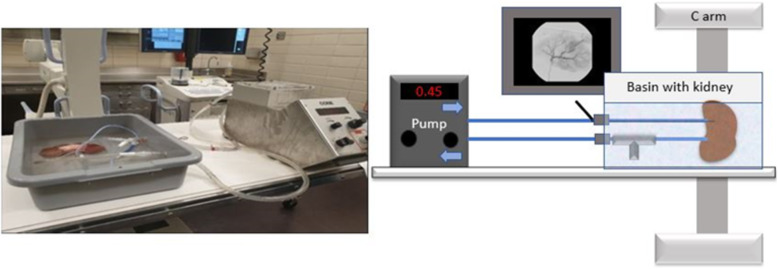
Objectives: To develop an ex- vivo perfusion flow model using a bovine kidney for future testing of embolic agents in an inexpensive and easy way.
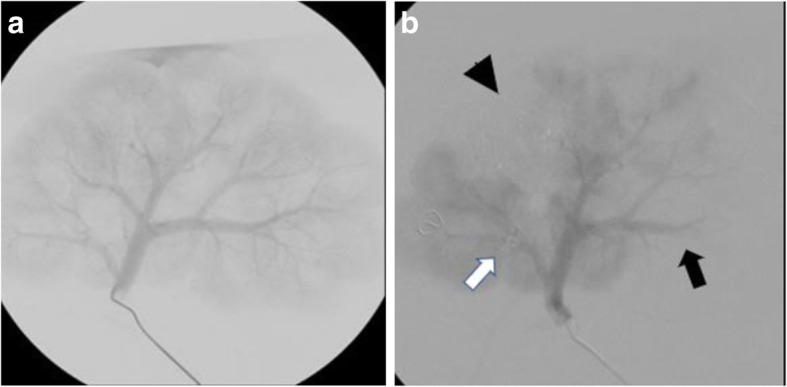
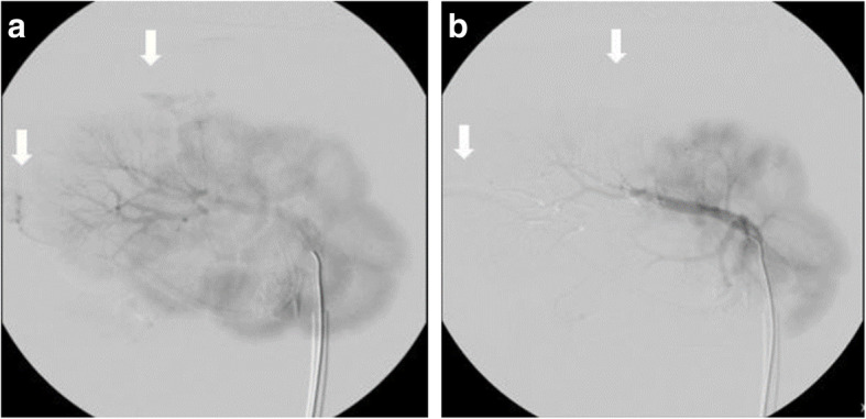
Methods: Six bovine adult kidneys were used for this study. Kidneys were cannulated and perfused via a roller pump. Three embolic agents, coils, Gelfoam, and a glue mixture of Histoacryl + Lipiodol, were deployed by targeting three secondary segmental arteries per kidney via a 5Fr catheter under fluoroscopic guidance. Cannulation time, success rate of segmental artery selection and embolic agent deployment, total operational time, and fluoroscopy dose were recorded.
Results: Average kidney weight was 0.752 +/- 0.094 kg. All six bovine kidneys were successfully cannulated in 21.6 min +/- 3.0 min. Deployment of coils and glue was achieved in every case (12/12); however, Gelfoam injection was not successful in one instance (5/6, 83%). Coil deployment demonstrated no embolic effect while Gelfoam and glue injections demonstrated decreased distal contrast filling post-embolization. Mean dose area product was 12.9 ± 1.8 Gy·cm2, fluoroscopy time was 10 ± 4 min and operational time was 27 ± 8 min.
Conclusions: We describe the creation of an ex vivo bovine kidney flow model for the preclinical evaluation of different embolic materials. The flow model can be modified to provide extensive bench testing and it is a promising tool for hands -on training in basic and advanced embolization techniques .
Keywords: Bovine kidney; Embolization; Flow model; Fluoroscopy; Perfusion.
Conflict of interest statement
None of the authors have any conflicts of interest to disclose.
Figures
References
-
- Ben-Menachem Y, Coldwell DM, Young JWR et al (1991) Hemorrhage associated with pelvic fractures: causes, diagnosis, and emergent management. Am J Roentgenol. 10.2214/ajr.157.5.1927786 - PubMed
LinkOut - more resources
Full Text Sources
Other Literature Sources