. 2021 Apr 26:39:167-198.
doi: 10.1146/annurev-immunol-110119-091711. Epub 2021 Feb 3.

Group 2 Innate Lymphoid Cells: Team Players in Regulating Asthma

Affiliations

Group 2 Innate Lymphoid Cells: Team Players in Regulating Asthma

Noe Rodriguez-Rodriguez et al. Annu Rev Immunol. .

Abstract

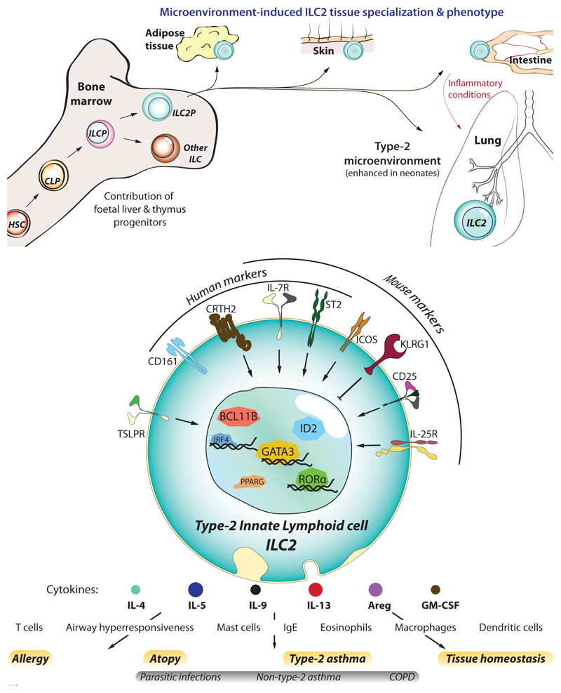
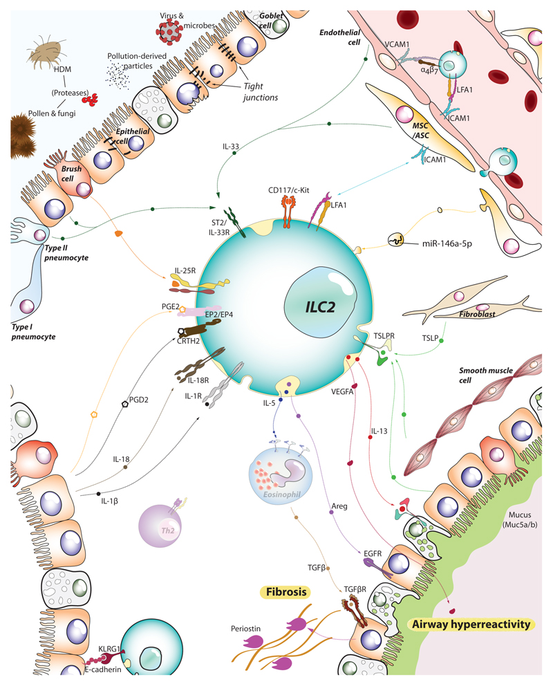
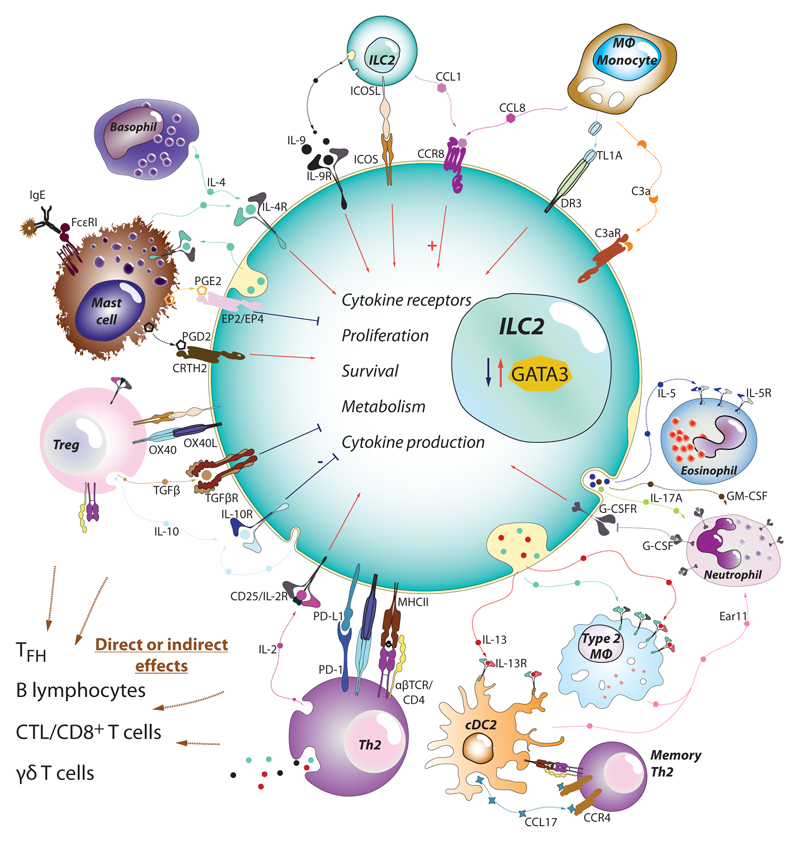
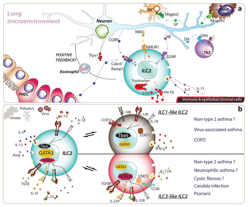
Type 2 immunity helps protect the host from infection, but it also plays key roles in tissue homeostasis, metabolism, and repair. Unfortunately, inappropriate type 2 immune reactions may lead to allergy and asthma. Group 2 innate lymphoid cells (ILC2s) in the lungs respond rapidly to local environmental cues, such as the release of epithelium-derived type 2 initiator cytokines/alarmins, producing type 2 effector cytokines such as IL-4, IL-5, and IL-13 in response to tissue damage and infection. ILC2s are associated with the severity of allergic asthma, and experimental models of lung inflammation have shown how they act as playmakers, receiving signals variously from stromal and immune cells as well as the nervous system and then distributing cytokine cues to elicit type 2 immune effector functions and potentiate CD4+ T helper cell activation, both of which characterize the pathology of allergic asthma. Recent breakthroughs identifying stromal- and neuronal-derived microenvironmental cues that regulate ILC2s, along with studies recognizing the potential plasticity of ILC2s, have improved our understanding of the immunoregulation of asthma and opened new avenues for drug discovery.

Keywords: alarmin; allergy; asthma; innate lymphoid cells; neuroimmunity; type 2 immunity.


Figures

Figure 1
Figure 1. ILC2 development and identification.
(a) Like other haematopoietic cells, ILC2s derive from haematopoietic stem cells (HSC) that differentiate into common lymphoid progenitors (CLP), and further commit towards the innate lymphocyte lineage as ILC progenitors (ILCP), and ILC2 progenitors (ILC2P). Though ILC2s first develop in foetal tissues, a significant fraction of them arise during the neonatal period whereupon they colonise most peripheral tissues (skin, intestine, lung, and visceral adipose tissue – VAT). Here they becom resident lymphocytes and adapt to the tissue microenvironment by acquiring a specialized genetic program tailored to each tissue. In particular, at least half of the ILC2s residing in the lung are from neonatal origin, a period where lungs display a strong type-2-biased microenvironment and allergic sensitisation likely occurs. However, under inflammatory conditions such as parasite infection or allergic reactions, the lung receives a transient ingress of ILC2s from the small intestine, which are characterized by increased sensitivity to IL-25 and higher expression of KLRG1, that contribute to lung inflammation. (b) ILC2s can be identified by their lack of lineage specific markers (CD3, CD4, CD8a, CD11b, CD11c, CD19, FcεRI, Ly6C, Ly6G, NK1.1/CD56, TCRαβ, TCRγδ, and Ter119), and expression of IL-7Rα (CD127) and high levels of the transcription factor GATA3. In addition, other molecules can serve to identify them in humans (i.e. CD161, CRTH2, and ST2) and mice (ST2, CD25, KLRG1, ICOS). Despite also providing contact-dependent signals, the foremost contribution of ILC2s to allergy and asthma is their overwhelming capacity to rapidly produce type-2 cytokines upon stimulation by cytokines/alarmins (IL-25, IL33, TSLP). In turn, ILC2-derived cytokines orchestrate the spectrum of immune effector cells that mediate the immunopathology of asthma, such as eosinophils and mast cells.
Figure 2
Figure 2
ILC2s sense the tissue microenvironment and kick-off the type-2 immune response. As tissue resident lymphocytes, ILC2s are strategically positioned in the mucosal barrier sites, poised to monitor the release of alarmins by epithelial, endothelial and stromal cells. Epithelial cells, brush cells, multipotent and adventitial stromal cells (MSC/ASC) and pneumocytes (together with immune cells such as alveolar macrophages and dendritic cells) can detect allergens and irritants and, in turn, release IL-33, IL-25 and TSLP. Together with IL-1β and IL-18 liberated by dying cells, these alarmins stimulate ILC2s to secrete IL-5, IL-13, amphiregulin (Areg), and vascular endothelial factor A (VEGFA), that engage their respective receptors in epithelial cells, goblet cells and smooth muscle cells amongst others. The activation of these cells leads to airway hyperresponsiveness, mucus production and fibrosis, cardinal pathophysiological features of asthma and allergy. Endothelial cells also respond to the local inflammation and support further recruitment of ILC2s and other immune effector cells from the blood and distal tissues (e.g. intestine), boosting the inflammatory process.
Figure 3
Figure 3
ILC2s as central playmakers within the immune cell network in asthma. Since their discovery, ILC2s have been found to directly interact with most of the immune cells participating in asthma and allergy. ILC2s integrate the information provided by alarmins with the signals derived from other innate leukocytes such as basophils, neutrophils, macrophages and mast cells. Cytokines (IL-2, IL-4, IL-9, IL-10…), chemokines (CCL1, CCL8), ligand-receptor interactions (ICOS-ICOSL), lipid mediators (PGD2, PGE2, PGI2, LTD4…) and other soluble factors will stimulate or suppress ILC2 expression of alarmin receptors, proliferation, survival, transcriptional factor levels (e.g. GATA3), and cytokine production. The net effect is the capacity of ILC2s to activate and positively modulate T cells, macrophages, dendritic cells into becoming type-2 immune cells (Th2, M2 or cDC2 respectively). In parallel, ILC2s also provide support for mast cells, eosinophils and neutrophils, effectors that will contribute extensively to the pathology associated with asthma
Figure 4
Figure 4. Environmental and microenvironmental cues leading to typical and atypical ILC2s in asthma.
(a) Recently, researchers have discovered that ILC2s express many receptors for neurotransmitters and neuropeptides, as well as exceptionally high levels of certain enzymes implicated in the synthesis of neurotransmitters. Both sympathetic and vagal sensory neurons project their afferent fibres within lung parenchyma. In particular, transient-receptor-potential-cation channel-subfamily-V-member-1-expressing nociceptive neurons (Trpv1+), which specialize in the detection of noxious stimuli from the environment (e.g. irritants, extreme temperatures, tissue damage), appear to play key roles in promoting airway hyperresponsiveness. Besides activating type-2 classical dendritic cells (cDC2) and mast cells through the production of Substance P (SP), and promoting type-2 T helper cell differentiation through the release of dopamine, lung afferent neurons release many peptides and biogenic amines which directly target ILC2s, which reside in close proximity of neural fibres. Neuromedin U (NMU) promotes ILC2 expansion and Th2 cytokine production while norepinephrine (NE) and calcitonin gene related peptide (CGRP) curb the activation of ILC2s. Furthermore, unusually for an immune cell, ILC2s can secrete CGRP and express the enzymatic machinery necessary to produce serotonin (5-TH). Murine models have shown that inhibition or deletion of nociceptive neurons prevents or decreases the severity of allergic immune responses and asthma, suggesting that modulation of neuro-immune units can represent a targetable treatment approach. (b) ILC2s are characterized by the expression of cardinal type-2 cytokines (i.e. IL-4, IL-5, IL-9, IL-13 and Areg) and the transcription factor, GATA3. Nonetheless, ILC2s exhibit a remarkable ability to adapt their biology to match that of their environment. This reversible plasticity allows ILC2s to acquire ILC1- and ILC3-like features such as specific expression of Tbet and RORγt and IFN-γ and IL-17A, respectively. Indeed, the changes induced by IL-1β and IL-12, in the case of ILC1-like ILC2s, or by IL-1β, IL-23 and TGFβ for ILC3-like ILC2s suggest a profound modulation in the transcriptional signature. Both human and murine ILC2s present this capacity in vitro, but we are only starting to understand the prevalence of this process in vivo. Interestingly, the plasticity of ILC2s provides a plausible hypothesis to understand how ILC2s may participate and promote COPD and non-type-2 asthma that are characterised by type-1 and type-3 immunity respectively.

References

    1. Croisant S. Epidemiology of asthma: prevalence and burden of disease. Adv Exp Med Biol. 2014;795:17–29. - PubMed
    1. Koczulla AR, Vogelmeier CF, Garn H, Renz H. New concepts in asthma: clinical phenotypes and pathophysiological mechanisms. Drug Discov Today. 2017;22:388–96. - PubMed
    1. Robinson DS, Hamid Q, Ying S, Tsicopoulos A, Barkans J, Bentley AM, Corrigan C, Durham SR, Kay AB. Predominant TH2-like bronchoalveolar T-lymphocyte population in atopic asthma. N Engl J Med. 1992;326:298–304. - PubMed
    1. Barlow JL, Bellosi A, Hardman CS, Drynan LF, Wong SH, Cruickshank JP, McKenzie AN. Innate IL-13-producing nuocytes arise during allergic lung inflammation and contribute to airways hyperreactivity. J Allergy Clin Immunol. 2012;129:191–8.:e1-4. - PubMed
    1. Halim TY, Krauss RH, Sun AC, Takei F. Lung natural helper cells are a critical source of Th2 cell-type cytokines in protease allergen-induced airway inflammation. Immunity. 2012;36:451–63. - PubMed

Publication types