. 2021 Apr 1;32(7):622-633.
doi: 10.1091/mbc.E20-06-0394. Epub 2021 Feb 3.

The IgG3 subclass of β1-adrenergic receptor autoantibodies is an endogenous biaser of β1AR signaling

Affiliations

The IgG3 subclass of β1-adrenergic receptor autoantibodies is an endogenous biaser of β1AR signaling

Maradumane L Mohan et al. Mol Biol Cell. .

Abstract

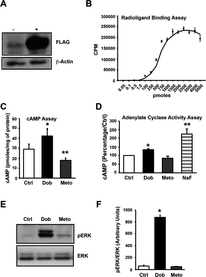
Dysregulation of immune responses has been linked to the generation of immunoglobulin G (IgG) autoantibodies that target human β1ARs and contribute to deleterious cardiac outcomes. Given the benefits of β-blockers observed in patients harboring the IgG3 subclass of autoantibodies, we investigated the role of these autoantibodies in human β1AR function. Serum and purified IgG3(+) autoantibodies from patients with onset of cardiomyopathy were tested using human embryonic kidney (HEK) 293 cells expressing human β1ARs. Unexpectedly, pretreatment of cells with IgG3(+) serum or purified IgG3(+) autoantibodies impaired dobutamine-mediated adenylate cyclase (AC) activity and cyclic adenosine monophosphate (cAMP) generation while enhancing biased β-arrestin recruitment and Extracellular Regulated Kinase (ERK) activation. In contrast, the β-blocker metoprolol increased AC activity and cAMP in the presence of IgG3(+) serum or IgG3(+) autoantibodies. Because IgG3(+) autoantibodies are specific to human β1ARs, non-failing human hearts were used as an endogenous system to determine their ability to bias β1AR signaling. Consistently, metoprolol increased AC activity, reflecting the ability of the IgG3(+) autoantibodies to bias β-blocker toward G-protein coupling. Importantly, IgG3(+) autoantibodies are specific toward β1AR as they did not alter β2AR signaling. Thus, IgG3(+) autoantibody biases β-blocker toward G-protein coupling while impairing agonist-mediated G-protein activation but promoting G-protein-independent ERK activation. This phenomenon may underlie the beneficial outcomes observed in patients harboring IgG3(+) β1AR autoantibodies.


Figures

FIGURE 1:
FIGURE 1:
Generation and characterization of stable HEK cell line expressing human β1-adrenergic receptors (β1AR). (A) Parental HEK 293 cells and HEK 293 cells overexpressing FLAG-human-β1AR (HEK-β1AR) were lysed with NP-40 lysis buffer, and cell lysates (50 μg each) were subjected to SDS–PAGE and immunoblotted with anti-FLAG antibody. The blots were stripped and immunoblotted with anti–β-actin antibody as loading control. (B) Isolated plasma membranes from HEK-β1AR cells were used to perform the receptor-binding assay using 125I-cyanopindolol to show the expression of receptors on the cell membranes and binding curve establish very high expression of the receptors (n = 3). (C) HEK-β1AR cells were serum starved for 4 h and stimulated with 10 μM dobutamine (Dob) or 10 μM metoprolol (Meto) for 10 min. The cells were lysed and cAMP generation was measured using a cAMP assay kit. Bar graphs represent cumulative data (n = 3 independent experiments, and each experiment is performed in triplicate). * p ≤ 0.05 Ctrl vs. Dob, ** p ≤ 0.01 Meto vs. Dob/Ctrl. (D) Isolated membranes from HEK-β1AR cells were used to perform the adenylate cyclase (AC) assay in vitro to assess the function of the expressed receptors in the presence of Dob or Meto. The amount of cAMP generated by control is expressed as 100%, and percent changes in the generation of cAMP in treatments are shown (n = 3). * p ≤ 0.05 Ctrl vs. Dob, ** p ≤ 0.001 NaF vs. Ctrl/Dob/Meto. (E) HEK-β1AR cells were serum starved and stimulated with Dob or Meto. The cell lysates were subjected to Western immunoblotting with anti–phospho-ERK. The blots were stripped and immunoblotted with anti-ERK antibody as loading control. (F) Cumulative densitometric data are presented as bar graphs (n = 3). * p ≤ 0.0001, Dob vs. Ctrl/Meto.
FIGURE 2:
FIGURE 2:
Autoantibody (AAb)-positive human serum alters β1AR function. (A) HEK-β1AR cells were serum starved and treated with human serum negative for the IgG3 class of AAb (IgG3[−]) or positive for the IgG3 class of AAb (IgG3[+]) for 10 min. The cAMP was measured (n = 3–4 independent patient serum samples). * p ≤ 0.01 IgG3(−) vs. Ctrl/IgG3(+). (B) HEK-β1AR cells were serum starved, pretreated with IgG3(−) human serum for 30 min, and stimulated with dobutamine (Dob) or metoprolol (Meto). The cAMP was measured (n = 3 independent patients). * p ≤ 0.05 Meto vs. Ctrl. (C) HEK-β1AR cells were serum starved, pretreated with IgG3(+) human serum, and stimulated with Dob or Meto. The cAMP was measured (n = 4). * p ≤ 0.05 Meto vs. Ctrl/Dob. (D) Isolated membranes from HEK-β1AR cells were pretreated with IgG3(−) or IgG3(+) human serum, and AC activity was determined in the presence of Dob or Meto. The amount of cAMP generated by control is expressed as 100%. Percent changes in generation of cAMP following treatments were calculated, and Δ-change compared with control is represented; n = 4 (no serum), 5 (IgG3[−] serum), or 7 (IgG3[+] serum). ** p ≤ 0.05 Dob human serum vs. Ctrl human serum, * p ≤ 0.01 Meto human serum vs. Dob human serum.
FIGURE 3:
FIGURE 3:
Autoantibodies (AAb) alter β1AR function. (A) HEK-β1AR cells were serum starved, pretreated with affinity-purified IgG3(−) AAb for 30 min, and stimulated with Dob or Meto. The cAMP was measured (n = 9). Δ-change compared with control is represented. * p ≤ 0.05 metoprolol (Meto) vs. dobutamine (Dob) (IgG3[−]). ** p ≤ 0.05 Meto vs. Dob (IgG3[+]). $ p ≤ 0.01 Meto (IgG3[+]) vs. Meto (IgG3[−]). (B) Isolated membranes from HEK-β1AR cells were pretreated with IgG3(−) purified IgG or IgG3(+) purified IgG, and AC activity was determined in the presence of Dob or Meto. The amount of cAMP generated by vehicle treatment (control) is expressed as 100%. Percent changes in generation of cAMP following treatments were calculated, and Δ-change compared with control is represented; n = 4 (no AAb), 5 (purified IgG3[−] AAb), or 7 (purified IgG3[+] AAb). ** p ≤ 0.05 Meto IgG3(+) purified IgG vs. Dob IgG3(+) purified IgG.
FIGURE 4:
FIGURE 4:
Autoantibodies (AAb) alter β-arrestin recruitment. (A) HTLA cells (HEK cells stably expressing tTA-dependent luciferase reporter and a β-arrestin2-TEV fusion gene) were transfected with the ADBR1-Tango (β1AR) gene, pretreated with human sera negative for IgG3 AAb or positive for IgG3 AAb for 30 min, subjected to β-arrestin recruitment assay, and compared with no IgG control (n = 8). * p ≤ 0.001 dobutamine (Dob) IgG3(+) serum vs. Dob IgG3(−) serum and Dob no IgG. (B) HTLA cells (HEK cells expressing tTA-dependent luciferase reporter and a β-arrestin2-TEV fusion gene) were transfected with the ADBR1-Tango (β1AR) gene pretreated with affinity-purified IgG3(−) or IgG3(+) AAb for 30 min, subjected to the β-arrestin recruitment assay, and compared with no IgG control (n = 8). * p ≤ 0.001 Dob IgG3(+) AAb vs. Dob IgG3(−) AAb or Dob no IgG.
FIGURE 5:
FIGURE 5:
Autoantibodies (AAb) alter membrane localization of β-arrestin. (A) HEK-β1AR cells transfected with plasmids containing the β-arrestin2-GFP fusion gene were plated on poly-l-lysine–coated coverslips, serum starved, pretreated with no IgG or affinity-purified IgG3(−) or IgG3(+) autoantibody, stimulated with Dob, fixed with 4% paraformaldehyde for 30 min, and mounted using prolong gold mountant with 4’,6-diamidino-2-phenylIndole (DAPI). The β-arrestin recruitment was assessed using confocal microscopy. Representative images of β-arrestin recruitment depicted by green fluorescence (bar = 10 μm). (B) Representative depiction of plot profile measurement of GFP fluorescence showing changes in fluorescence intensities from one edge to the other edge of the cell, which depicts the reduction of fluorescene intensity in the cytoplasm and increase at the plasma membrane following dobutamine (Dob treatment. (C) The relative ratio of plasma membrane (Mem) to cytoplasm (Cyt) intensity with Dob treatment in the presence of no IgG or IgG3(−) or IgG3(+) AAb (n = 3). * p<0.05 IgG3(+) vs. IgG3(−) AAb or no IgG. (D) Average changes (i.e,. clearance/loss) in cytoplasmic fluorescence intensity calculated over total fluorescence intensity of the cell reflecting β-arrestin recruitment, represented as percentage (∼ 30 cells/experiment [n = 3]). * p < 0.05 IgG3(+) AAb vs. no IgG or IgG3(−) AAb. (E) Percentage of cells showing effective β-arrestin recruitment following Dob in the presence of no IgG or IgG3(−) or IgG3(+) AAb (∼30 cells/experiment [n = 3]). *p < 0.05 IgG3(+) AAb vs. no IgG or IgG3(−) AAb.
FIGURE 6:
FIGURE 6:
Autoantibodies (AAb) alter β1AR signaling. (A) HEK-β1AR cells were serum starved for 4 h, pretreated with IgG3(−) or IgG3(+) human serum for 30 min, and stimulated with dobutamine (Dob) or metoprolol (Meto). The cell lysates were subjected to Western immunoblotting with anti–phospho-ERK antibody. The blots were stripped and immunoblotted with anti-ERK antibody as loading control. (B) Cumulative data for cells pretreated with IgG3(−) human serum (n = 3). (C) Cumulative data for cells pretreated with IgG3(+) human serum (n = 3). (D) HEK-β1AR cells were serum starved, pretreated with no IgG or affinity-purified IgG3(−) or IgG3(+) AAb and stimulated with Dob or Meto. The cell lysates were subjected to Western immunoblotting as above. (E) Cumulative data (n = 3). *p ≤ 0.05 Ctrl IgG3(−)/IgG3(+) vs. Ctrl no IgG. **p ≤ 0.01 Dob IgG3(−)/IgG3(+) vs. Dob no IgG. $ p ≤ 0.05 Meto IgG3(+) vs. Meto no IgG/IgG3(−).
FIGURE 7:
FIGURE 7:
Autoantibodies (AAb) alter β1AR function in human hearts. (A) Isolated membranes from donor human heart tissues were pretreated with no AAb, purified IgG3(−) AAb, or purified IgG3(+) AAb, and AC activity was determined in the presence of dobutamine (Dob) or metoprolol (Meto). The amount of cAMP generated by control is expressed as 100%. Percent changes in generation of cAMP following treatments were calculated, and Δ-change compared with control is represented (n = 4). *p ≤ 0.05 Dob IgG3(+) purified IgG vs. Dob IgG(−) purified IgG/no IgG. **p ≤ 0.005 Meto IgG3(+) purified IgG vs. Meto IgG3(−) purified IgG/no IgG. (B) AC activity determined as above. The Dob-mediated cAMP is expressed as percent over control. *p ≤ 0.05 IgG3(−) purified IgG vs. no IgG. **p ≤ 0.05 IgG3(+) purified IgG vs. IgG3(−) purified IgG/no IgG. (C) AC activity determined as above. The Meto-mediated cAMP is expressed as percent over control. *p ≤ 0.05 IgG3(+) purified IgG vs. IgG3(−) purified IgG/no IgG.
FIGURE 8:
FIGURE 8:
Autoantibodies (AAb)-positive human serum does not alter β2AR signaling. (A) HEK 293 cells overexpressing FLAG-human-β2AR (HEK-β2AR) were serum starved for 4 h, pretreated with no IgG or IgG3(−) or IgG3(+) human serum for 30 min, stimulated with 10 μM isoproterenol (Iso) or 10 μM ICI for 10 min, and lysed with NP-40 lysis buffer, and cell lysates (50 μg each) were subjected to SDS–PAGE and immunoblotted with anti–phospho-β2AR antibody. The blots were stripped and immunoblotted with anti-FLAG antibody as loading control. The cell lysates were subjected to Western immunoblotting with anti–phospho-β2AR. The blots were stripped and immunoblotted with anti-FLAG antibody as loading control. (B) Cumulative data for cells pretreated with no IgG3 human serum (n = 3). *p ≤ 0.01 Iso vs. Ctrl/ICI. (C) HEK-β2AR cells were serum starved, pretreated with IgG3(−) human serum, and stimulated with Iso or ICI. The cell lysates were subjected to Western immunoblotting as above (n = 3). *p ≤ 0.01 Iso vs. Ctrl/ICI. (D) HEK-β2AR cells were serum starved, pretreated with IgG3(+) human serum, and stimulated with Iso or ICI. The cell lysates were subjected to Western immunoblotting as above (n = 4). *p ≤ 0.01 Iso vs. Ctrl/ICI. (E) HEK-β2AR cells were serum starved, pretreated with no IgG or IgG3(−) or IgG3(+) human serum, and stimulated with Iso and ICI. The cell lysates were subjected to Western immunoblotting with anti–phospho-ERK. The blots were stripped and immunoblotted with anti-ERK antibody as loading control. (F) Cumulative data for cells pretreated with no IgG human serum (n = 3). *p ≤ 0.01 Iso vs. Ctrl/ICI. (G) Cumulative data for cells pretreated with IgG3(−) human serum (n = 3). *p ≤ 0.05 Iso vs. Ctrl/ICI. (H) Cumulative data for cells pretreated with IgG3(+) human serum (n = 4). *p ≤ 0.05 Iso vs. Ctrl/ICI.
FIGURE 9:
FIGURE 9:
Illustration depicting signaling mechanism of IgG3(+) autoantibodies (AAb) generated against human β1AR. (A) Agonist-mediated β1AR signaling. (B) Antagonist-mediated β1AR signaling. (C) Agonist-mediated β1AR signaling modulated by IgG3(+) AAb. (D) Antagonist-mediated β1AR signaling modulated by IgG3(+) AAb.

Similar articles

Cited by

References

    1. Baba A (2010). Targeted autoantibodies in apheresis treatment against severe heart failure. Jpn J Apheresis 29, 187–193.
    1. Bornholz B, Weidtkamp-Peters S, Schmitmeier S, Seidel CA, Herda LR, Felix SB, Lemoine H, Hescheler J, Nguemo F, Schafer C, et al. (2013). Impact of human autoantibodies on beta1-adrenergic receptor conformation, activity, and internalization. Cardiovasc Res 97, 472–480. - PMC - PubMed
    1. Bristow MR (1997). Mechanism of action of beta-blocking agents in heart failure. Am J Cardiol 80, 26L–40L. - PubMed
    1. Carr R 3rd, Schilling J, Song J, Carter RL, Du Y, Yoo SM, Traynham CJ, Koch WJ, Cheung JY, Tilley DG, et al. (2016). beta-Arrestin-biased signaling through the beta2-adrenergic receptor promotes cardiomyocyte contraction. Proc Natl Acad Sci USA 113, E4107–E4116. - PMC - PubMed
    1. Christ T, Wettwer E, Dobrev D, Adolph E, Knaut M, Wallukat G, Ravens U (2001). Autoantibodies against the beta1 adrenoceptor from patients with dilated cardiomyopathy prolong action potential duration and enhance contractility in isolated cardiomyocytes. J Mol Cell Cardiol 33, 1515–1525. - PubMed

Publication types

LinkOut - more resources