. 2021 Feb 3;17(2):e1009223.
doi: 10.1371/journal.ppat.1009223. eCollection 2021 Feb.

The leucine-rich repeats in allelic barley MLA immune receptors define specificity towards sequence-unrelated powdery mildew avirulence effectors with a predicted common RNase-like fold

Affiliations

The leucine-rich repeats in allelic barley MLA immune receptors define specificity towards sequence-unrelated powdery mildew avirulence effectors with a predicted common RNase-like fold

Saskia Bauer et al. PLoS Pathog. .

Abstract

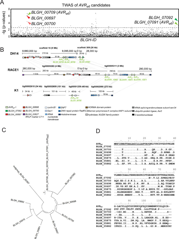
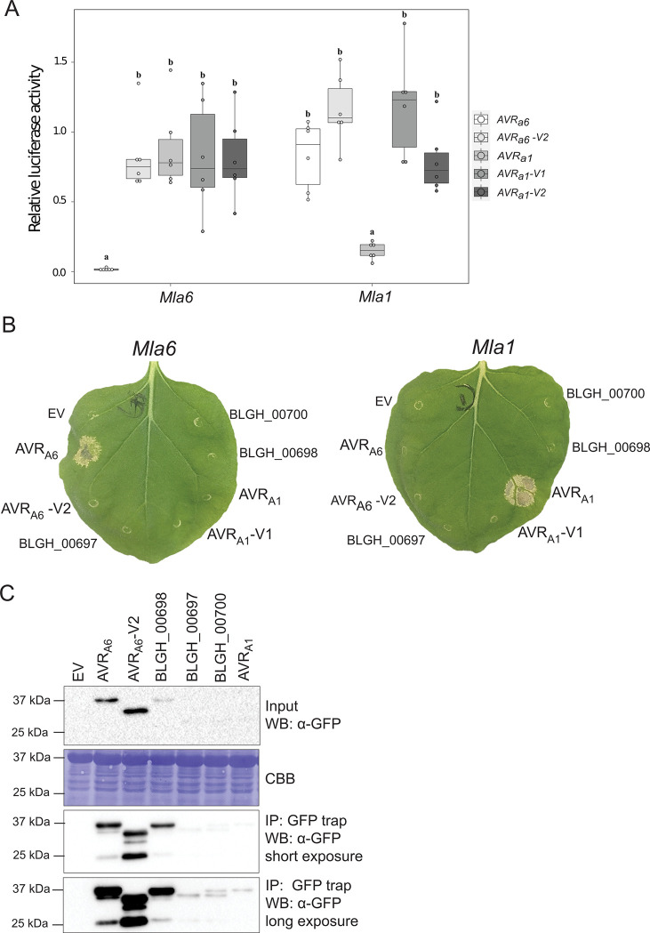
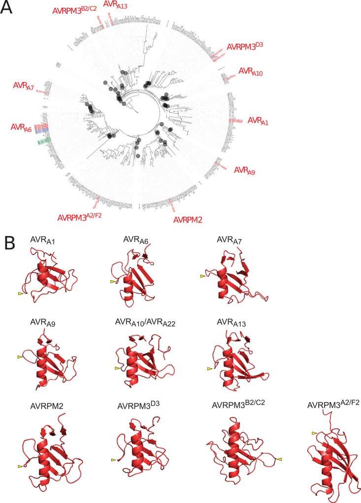
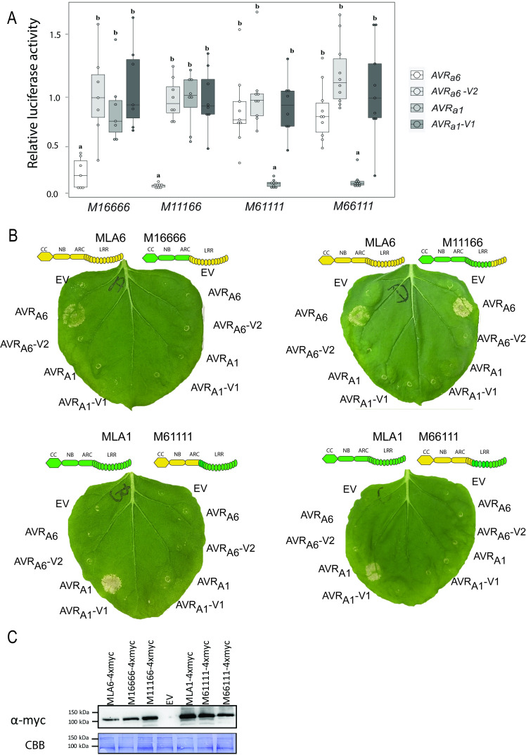
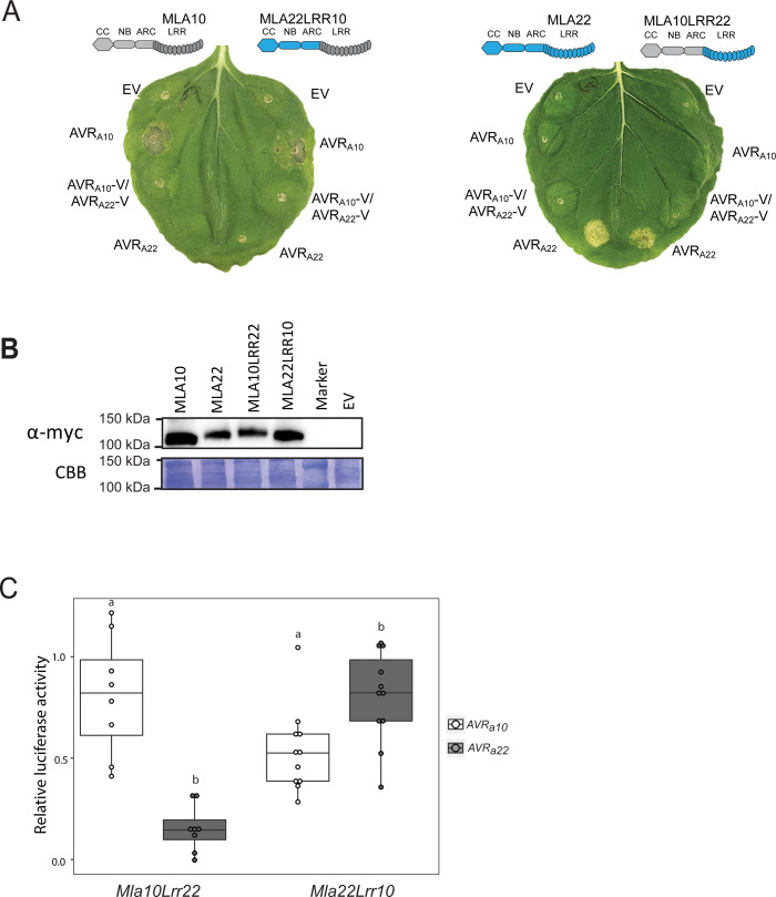
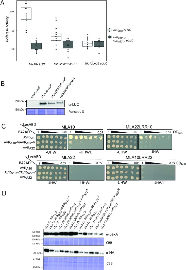
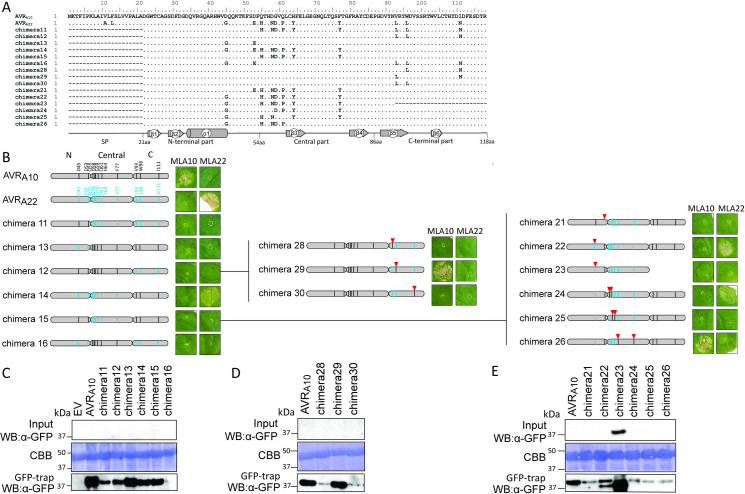
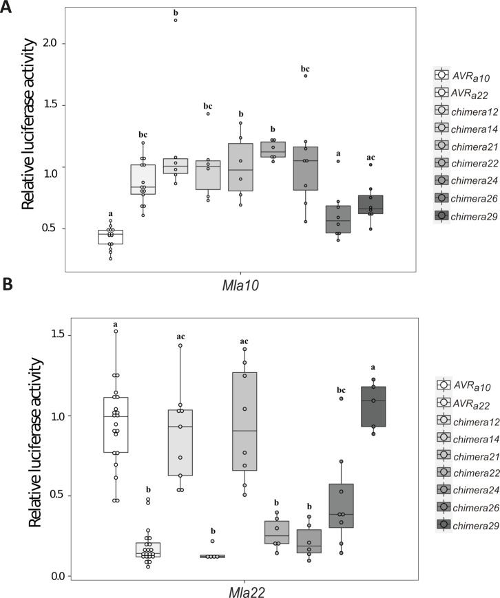
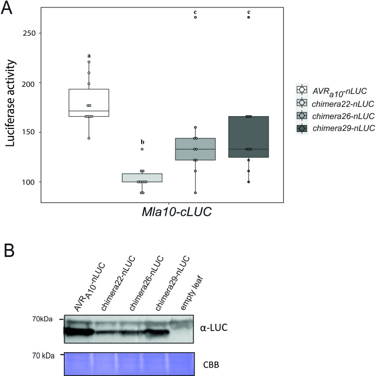
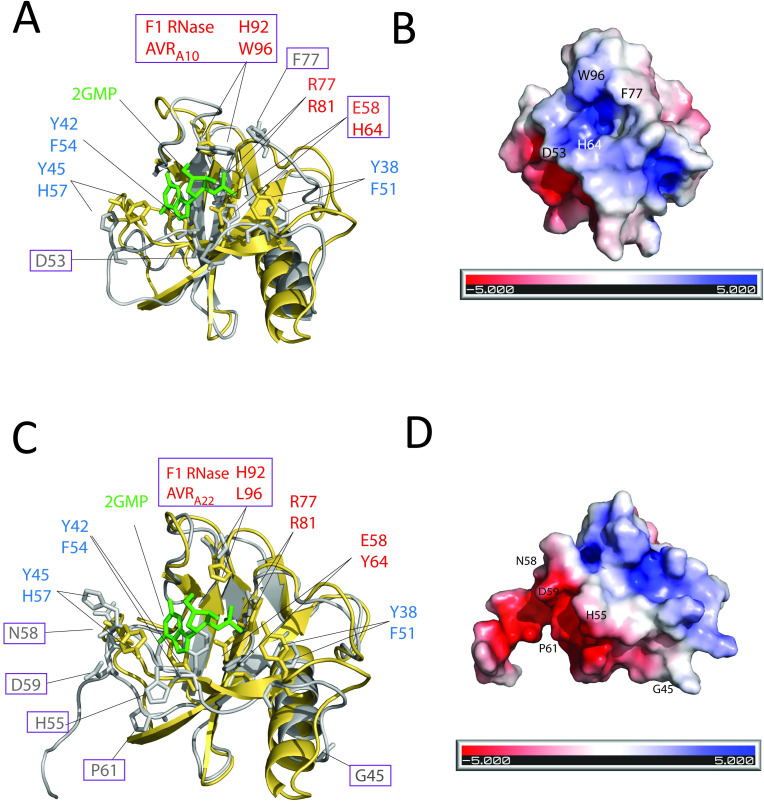
Nucleotide-binding domain leucine-rich repeat-containing receptors (NLRs) in plants can detect avirulence (AVR) effectors of pathogenic microbes. The Mildew locus a (Mla) NLR gene has been shown to confer resistance against diverse fungal pathogens in cereal crops. In barley, Mla has undergone allelic diversification in the host population and confers isolate-specific immunity against the powdery mildew-causing fungal pathogen Blumeria graminis forma specialis hordei (Bgh). We previously isolated the Bgh effectors AVRA1, AVRA7, AVRA9, AVRA13, and allelic AVRA10/AVRA22, which are recognized by matching MLA1, MLA7, MLA9, MLA13, MLA10 and MLA22, respectively. Here, we extend our knowledge of the Bgh effector repertoire by isolating the AVRA6 effector, which belongs to the family of catalytically inactive RNase-Like Proteins expressed in Haustoria (RALPHs). Using structural prediction, we also identified RNase-like folds in AVRA1, AVRA7, AVRA10/AVRA22, and AVRA13, suggesting that allelic MLA recognition specificities could detect structurally related avirulence effectors. To better understand the mechanism underlying the recognition of effectors by MLAs, we deployed chimeric MLA1 and MLA6, as well as chimeric MLA10 and MLA22 receptors in plant co-expression assays, which showed that the recognition specificity for AVRA1 and AVRA6 as well as allelic AVRA10 and AVRA22 is largely determined by the receptors' C-terminal leucine-rich repeats (LRRs). The design of avirulence effector hybrids allowed us to identify four specific AVRA10 and five specific AVRA22 aa residues that are necessary to confer MLA10- and MLA22-specific recognition, respectively. This suggests that the MLA LRR mediates isolate-specific recognition of structurally related AVRA effectors. Thus, functional diversification of multi-allelic MLA receptors may be driven by a common structural effector scaffold, which could be facilitated by proliferation of the RALPH effector family in the pathogen genome.


Conflict of interest statement

The authors have declared that no competing interests exist.

Figures

Fig 1
Fig 1. Identification of BLGH_00709 (CSEP0254) as one of the top ranking AVRa6 candidates by association of Bgh AVRa profiles on Mla6 near-isogenic lines with transcript polymorphisms.
(A) Manhattan plot summarizing the gene-wise association results for candidate AVRa6. The x axis represents the Bgh DH14 genes, sorted by Bgh gene ID; the y axis shows −lg of p-values for all genes with at least one nonsynonymous coding SNP, indels as well as presence or absence of transcripts. CSEPs with a p < 0.018 (dotted line) are depicted by arrowheads. The candidate AVRa6 gene copies BLGH_00709 (CSEP0254) and BLGH_07091 are designated in the plot with bright green arrowheads. BLGH_07092 is designated by a dark green arrowhead. The other candidates BLGH_00697 (CSEP0058) and BLGH_00700 are depicted with a dark red and a bright red arrowhead, respectively. (B) Schematic illustration of the chromosomal regions harboring the AVRa6 candidate BLGH_00709 and its paralogues and family members with corresponding gene IDs in the genomes of Bgh isolates DH14, RACE1, and K1. All CSEPs are depicted by arrows. (C) Phylogeny of CSEP family 8 containing AVRA6, which can be divided into clade 1 (BLGH_00709, BLGH_07092, BLGH_00700, BLGH_00698, BLGH_00697) and clade 2 (BLGH_05882, BLGH_05875, BLGH_05881), based on the protein sequences excluding the signal peptide and using BLGH_05397 as an outgroup. (D) Protein sequence alignment of AVRA6 and CSEP family 8 members including their respective signal peptides.
Fig 2
Fig 2. Mla6 and AVRa6 co-expression in barley protoplasts and N. benthamiana causes a specific cell death response.
(A) Barley cv. Golden Promise protoplasts were transfected with pIPKb002 vectors containing cDNAs of Mla6 or Mla1 and either an empty vector (EV), AVRa6, AVRa6-V2, AVRa1, AVRa1-V1, or AVRa1-V2 variants lacking their respective signal peptides together with a pUBI:Luciferase construct. The LUC activity relative to the EV sample was measured as a proxy for cell death 16 h post transfection. Box plot diagrams show median of the relative LUC activity of six independent transfections, which are represented by dots, while the box shows the interquartile range. Significant differences between samples were analyzed using non-parametric Kruskal-Wallis (KW) analysis followed by a Dunn’s test. Calculated KW p-values are as follows: Mla6: p = 0.007146; Mla1: p = 0.0007392. Samples labeled with identical letters did not differ significantly (p < 0.05) in the Dunn’s test for the corresponding Mla variant. (B) cDNAs of clade 1 AVRa6 family members BLGH_00698, BLGH_00697, BLGH_00700, AVRa6, and AVRa1 variants were expressed without their respective signal peptides and stop codons and with a C-terminal mYFP fusion under the control of a 35S promotor in N. benthamiana. The effectors were co-expressed with Mla1 and Mla6 cDNAs fused C-terminally with a 4xmyc tag under the control of a 35S promotor. Cell death was scored five days post infiltration and Figures show a representative of at least 15 co-transformations. (C) Protein levels of AVRA1-mYFP, AVRA6-mYFP, AVRA6-V2-mYFP, BLGH_00698-mYFP, BLGH_00697-mYFP and BLGH_00700-mYFP. Samples for total protein extraction were harvested two days post infiltration. mYFP fusion proteins were enriched by an GFP-Trap. Proteins were separated using 10% or 12% polyacrylamide gels and proteins were detected using α-GFP and α-myc western blotting (WB). IP = immunoprecipitation. CBB = Coomassie Brilliant Blue.
Fig 3
Fig 3. Bgh AVRA and Bgt AVRPM effectors are sequence-unrelated but exhibit predicted structural similarity to RNases.
(A) Maximum likelihood phylogeny including all predicted CSEPs from B. graminis formae speciales poae, lolium, avenae, tritici 96224, hordei DH14, secalis S1459, triticale T1-20, and dactylidis. Depicted in red are the BLGH-IDs of all so far isolated Bgh AVRA and Bgt AVRPM effectors. Depicted in blue are the clade-1 family members of AVRA6, while the clade-2 family members are colored in green. CSEP clades that were collapsed (grey circles) to improve legibility of the tree do not include AVR members and are indicated by grey circles. (B) Structural prediction of isolated AVRAs and AVRPM by IntFOLD version 5.0 in red (p-values: AVRA1 = 4.888e-4 most similar to PDB IDs 5gy6, 3who and 1rds, AVRA6 = 3.293e-5 to 6fmb, AVRA7 = 2.114e-4 to PDB ID 5gy6, AVRA9 = 1.18e-5 most similar to PDB IDs 6fmb, 3who, and 1ch0, AVRA10 = 9.759e-5 most similar to PDB ID 1fusa and to 3whoa, AVRA13-1 = 7,359e-7 most similar to 6fmb, AVRPM2 = 8.741e-9 most similar to 6fmb, 1chOA, and 1rmsA, AVRPM3D3 = 8.82e-5 most similar to PDB 6fmb and 5gy6A, AVRPM3A2/F2 = 1.145e-5 to 3ub1A2, AVRPM3B2/C2 = 7.079e-2, no structural similarities predicted). Yellow arrow depicts relative position of the characteristic RALPH intron in effector structures.
Fig 4
Fig 4. Recombinant AVRA6, AVRA10, and AVRA13 effector proteins do not exhibit ribonuclease activity.
To test for ribonuclease activity, heterologous AVRA6, AVRA10, and AVRA13 proteins, purified upon expression of the respective genes in E. coli, or T1 RNase were co-incubated with (A) denatured HvRNA and (B) native rabbit rRNA always. All samples were separated on non-denaturing 2% agarose gels (top panels) and analysed on a Bioanalyzer (lower panels) to check for RNA degradation.
Fig 5
Fig 5. Specific recognition of AVRA6 and AVRA1 by MLA1/MLA6 chimeric constructs in planta.
(A) Barley cv. Golden Promise protoplasts were transfected with a LUC reporter construct and pIPKb002 vectors containing cDNAs of AVRa6, AVRa6-V2, AVRa1, AVRa1-V1 or an empty vector (EV) together with vectors harboring intron-containing DNA of receptor chimeras M16666, M11166, M61111, or M66111 under the control of a pZmUBI promotor. Transfections were performed at least seven times independently. Significant differences between samples were analyzed using non-parametric Kruskal-Wallis (KW) analysis followed by a Dunn’s test. Calculated KW p-values are as follows: M16666: p = 0.001883; M11166: p = 0.000559, M61111: p = 0.0001582, M66111: p = 1.658e-05. Samples labeled with identical letters did not differ significantly (p < 0.05) in the Dunn’s test for the corresponding Mla variant. (B) Transient transformation of N. benthamiana leaves with empty vector (EV) or cDNAs of AVRa6 or AVRa1 variants fused C-terminally with a mYFP tag together with Mla1 or Mla6 cDNAs or M16666, M11166, M61111, or M66111 intron-containing DNAs with a C-terminal 4xmyc fusion. All constructs were expressed from a 35S promotor. Figures show a representative of at least three independent co-transformations. (C) MLA-4xmyc proteins were extracted two days post infiltration and separated using a 10% polyacrylamide gels and detected using α-myc western blotting, CBB = Coomassie Brilliant Blue.
Fig 6
Fig 6. The LRR domains of MLA10 and MLA22 distinguish between AVRA10 and AVRA22.
(A) Transient transformation of Nicotiana benthamiana leaves with EV or cDNAs of AVRa10, AVRa10-V/AVRa22-V, and AVRa22 fused C-terminally with mYFP together with Mla10 or Mla22 cDNAs fused C-terminally with a 4xmyc tag. All constructs were expressed from a 35S promotor. Cell death was scored five days post infiltration (dpi) and Figures show a representative of at least 15 co-transformations. (B) Protein levels of MLA-4xmyc after total protein extraction from N. benthamiana leaves at two dpi. Proteins were separated on a 10% polyacrylamide gel and a detected using α-myc western blotting (WB) (C) Barley cv. Golden Promise protoplasts were transfected with a LUC reporter construct and pIPKb002 vectors containing cDNAs of AVRa10 or AVRa22 together with Mla10Lrr22 and Mla22Lrr10 chimeras. Transfections were performed at least eight times independently. Significant differences between samples were analyzed using non-parametric Kruskal-Wallis (KW) one-way analysis of variance. Calculated KW p-values are as follows: Mla10Lrr22: p = 0.0007775; Mla22Lrr10: p = 0.01654. Samples labeled with different letters differed significantly (p < 0.05).
Fig 7
Fig 7. The LRR domain of MLA10 accounts for the specific interaction with AVRA10 in planta and in yeast.
(A) Nicotiana benthamiana leaves were transformed transiently with vectors containing cDNAs of Mla10-cLUC, Mla10Lrr22-cLUC or Mla22Lrr10-cLUC together with vectors containing cDNAs of AVRa10-nLUC or AVRa10-V/ AVRa22-V nLUC lacking signal peptides (SPs), under the control of a 35S promoter. LUC activity was determined forty hours after transfection. The experiment was performed on at least three independent days with two to four replicates (independent set of plants) each day. Significant differences between AVRa10-nLUC or AVRa10-V/ AVRa22-V nLUC were analyzed using one-way Kruskal-Wallis (KW) analysis. Calculated KW p-values are as follows: Mla10: p = 0.0001491; Mla22Lrr10: p = 0.009035; Mla10Lrr22: p = 0.8079. Samples labeled with different letters differed significantly (p < 0.05). (B) Protein levels of MLA10-cLUC, MLA22LRR10-cLUC and MLA10LRR22-cLUC in N. benthamiana leaf extracts. Proteins were separated on a 8% SDS-PAGE gel and a detected using anti-LUC western blot (WB). (C) Yeast was co-transformed with cDNAs of N-terminal LexABD-fused MLA and N-terminal B42AD-fused AVRA variants. Growth on media lacking Leucine indicates association of respective proteins fused to AD (activation domain) and BD (Binding domain). (D) Protein levels of LexABD-MLA and B42AD-AVRA variants. Proteins were precipitated using an ammonium-acetate buffer and dissolved in a urea-SDS sample buffer before separation on a 10% or 12% polyacrylamide gel and detection by either α-LexA or α-HA WB.
Fig 8
Fig 8
Co-expression of AVRA10 and AVRA22 chimeras with MLA10 and MLA22 in N. benthamiana. (A) Protein sequence alignment of the AVRA10/AVRA22 chimeric construct. Grey boxes below sequences show the length of the N-terminal, the central and the C-terminal effector part. Dashes represent missing/deletion of effector parts and points designate identical amino acid residues (B) Co-expression of N. benthamiana with cDNAs of AVRa10, AVRa22, or chimeric AVRa10/AVRa22 constructs fused C-terminally with mYFP and Mla10 or Mla22 cDNAs fused C-terminally with a 4xmyc tag from the 35S promotor in N. benthamiana leaves. Cell death was scored five days post infiltration and Figures show a representative of at least seven co-transformations. (C-E) Protein levels of AVRA-mYFP and chimeric AVRA10/AVRA22mYFP variants after total protein extraction from N. benthamiana leaves at two dpi. Proteins were separated on a 12% polyacrylamide gel and detected by α-mYFP western blotting.
Fig 9
Fig 9. Co-expression of a selection of AVRA10/AVRA22 chimeras with MLA10 and MLA22 in barley protoplasts.
Barley protoplasts of cv. Golden Promise were co-transfected with a LUC reporter assay and pIPKb002 vectors containing cDNAs of AVRa10, AVRa22, chimera12, chimera14, chimera21, chimera22, chimera24, chimera26 or chimera29 without signal peptide or with an empty vector (EV) together with either (A) Mla10 or (B) Mla22 in pipkb002. Transfections were performed at least six times independently. Significant differences between samples were analyzed using non-parametric Kruskal-Wallis (KW) analysis followed by the Dunn’s test. Calculated KW p-value are as follows: Mla10: p = 1.439e-07, Mla22: p = 5.374e-11. Samples labeled with different letters differed significantly (p < 0.05) in the Dunn’s test.
Fig 10
Fig 10. AVRA10 amino acid residues that are responsible for specific recognition correlate with residues that interact with the MLA10 receptor.
(A) N. benthamiana plants were transformed transiently with vectors containing cDNAs of Mla10-cLUC together with cDNAs of AVRa10-nLUC without signal peptide, chimera22-nLUC, chimera26-nLUC or chimera29-nLUC under the control of a 35S promoter. LUC activity was determined 40 h after A. tumefaciens-mediated transformation. The experiment was performed on at least four independent days with two to four replicates (independent set of plants) each day. Significant differences between samples were analyzed using non-parametric Kruskal-Wallis (KW) analysis followed by the Dunn’s test. Calculated KW p-value = 5.03e-05. Samples labeled with different letters differed significantly (p < 0.05) in the Dunn’s test. (B) Protein levels of AVRA10-nLUC, chimera22-nLUC, chimera26-nLUC and chimera29-nLUC. Proteins were separated on a 8% SDS_PAGE gel and a detected using anti-LUC western blotting (WB).
Fig 11
Fig 11. The location of amino acid residues in AVRA10 and AVRA22 that determine MLA10 and MLA22 recognition specificities.
(A and C) show structural superimposition of the crystal structure of Fusarium moniliformis RNase F1 (yellow) and IntFOLD version 5.0 structural predictions of AVRA10 or AVRA22 (grey). Depicted in green is the F1 RNase ligand 2’-guanosine monophosphate (2’ GMP); residues of the F1 RNase catalytic triad and corresponding AVRA residues are depicted in red; residues of the F1 RNase RNA binding pocket and corresponding AVRA residues are shown in blue. The residues of AVRA10 and AVRA22 required for specific MLA10 and MLA22 recognition as determined in Figs 8–10 are framed with a purple rectangle. (B and D) Predicted electrostatic surface potential of the AVRA10 and AVRA22 effector surface calculated using Adaptive Poisson Boltzmann Solver (APBS) [67]. The residues of AVRA10 and AVRA22 required for specific MLA10 and MLA22 recognition as determined in Figs 8–10 are indicated. Below is a scale bar of the electrostatic potential (red = negative charge, white = neutral charge, blue = positive charge).

Similar articles

Cited by

References

    1. Han G-Z. Origin and evolution of the plant immune system. New Phytol. 2019;222: 70–83. 10.1111/nph.15596 - DOI - PubMed
    1. van der Burgh AM, Joosten MHAJ. Plant Immunity: Thinking Outside and Inside the Box. Trends Plant Sci. 2019;24: 587–601. 10.1016/j.tplants.2019.04.009 - DOI - PubMed
    1. Periyannan S, Milne RJ, Figueroa M, Lagudah ES, Dodds PN. An overview of genetic rust resistance: From broad to specific mechanisms. PLoS Pathog. 2017;13: e1006380–e1006380. 10.1371/journal.ppat.1006380 - DOI - PMC - PubMed
    1. Dyck PL, Kerber ER. Resistance of the Race-Specific Type. Cereal rusts. 1985;II.
    1. Flor HH. Inheritance of pathogenicity in Melampsora lini. Phytopathology; 1942. pp. 653–669.

Publication types

MeSH terms

Supplementary concepts

LinkOut - more resources