. 2021 Mar;25(6):2885-2899.
doi: 10.1111/jcmm.16319. Epub 2021 Feb 3.

Paeonol attenuates inflammation by confining HMGB1 to the nucleus

Affiliations

Paeonol attenuates inflammation by confining HMGB1 to the nucleus

Jifei Miao et al. J Cell Mol Med. 2021 Mar.

Abstract

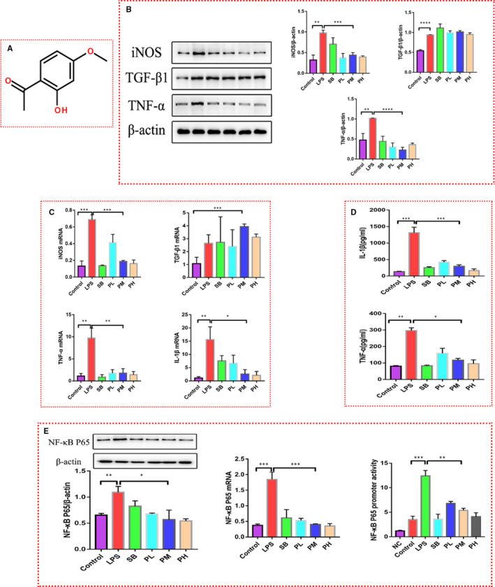
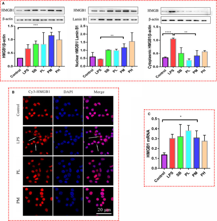
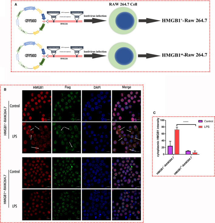
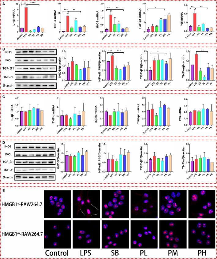
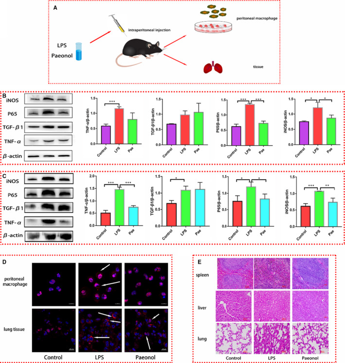
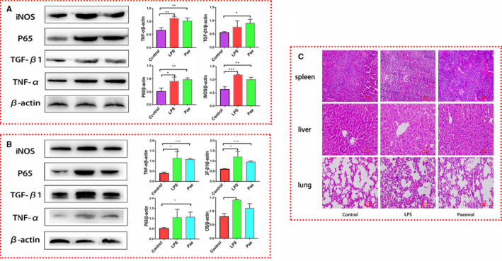
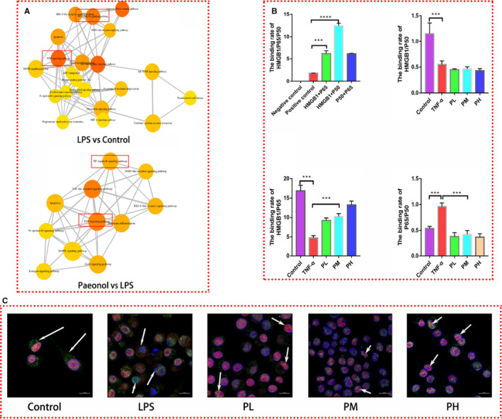
Inflammation is a biological process that exists in a large number of diseases. If the magnitude or duration of inflammation becomes uncontrolled, inflammation may cause pathological damage to the host. HMGB1 and NF-κB have been shown to play pivotal roles in inflammation-related diseases. New drugs aimed at inhibiting HMGB1 expression have become a key research focus. In the present study, we showed that paeonol (Pae), the main active component of Paeonia suffruticosa, decreases the expression of inflammatory cytokines and inhibits the translocation of HMGB1 induced by lipopolysaccharide (LPS). By constructing HMGB1-overexpressing (HMGB1+ ) and HMGB1-mutant (HMGB1m ) RAW264.7 cells, we found that the nuclear HMGB1 could induce an LPS-tolerant state in RAW264.7 cells and that paeonol had no influence on the expression of inflammatory cytokines in HMGB1m RAW264.7 cells. In addition, the anti-inflammatory property of paeonol was lost in HMGB1 conditional knockout mice, indicating that HMGB1 is a target of paeonol and a mediator through which paeonol exerts its anti-inflammatory function. Additionally, we also found that HMGB1 and P50 competitively bound with P65, thus inactivating the NF-κB pathway. Our research confirmed the anti-inflammation property of paeonol and suggests that inhibiting the translocation of HMGB1 could be a new strategy for treating inflammation.

Keywords: HMGB1; NF-κB; P65; inflammation; paeonol.


Conflict of interest statement

The authors declare that they have no conflicts of interest.

Figures

Figure 1
Figure 1
Paeonol attenuates the inflammation induced by LPS in RAW264.7 cells. A, The structure of paeonol is shown. B, iNOS, TNF‐α and TGF‐β1 levels in RAW264.7 cells treated with LPS, SB or paeonol were tested by using Western blotting. C, iNOS, TNF‐α, TGF‐β1 and IL‐1β levels were tested by using RT‐qPCR. D, IL‐1β and TNF‐α levels in cell culture supernatant were tested by using ELISA. E, Left: the protein level of P65 was tested by using Western blotting. Middle: the mRNA level of P65 was tested by using RT‐qPCR. Right: the promoter activity of P65 was tested by using a luciferase reporter assay. The bar graphs are representative of three independent experiments. *P < .05, ** P < .01 and *** P < .001
Figure 2
Figure 2
Paeonol inhibits the translocation of HMGB1 from the nucleus to the cytoplasm. A, Total protein (left), nuclear protein (middle) and cytoplasmic protein (right) were extracted. The level of HMGB1 in each fraction was tested by using Western blotting. B, HMGB1 was stained with Cy3 (red), and nuclei were stained with DAPI (blue). HMGB1 was distributed in the cytoplasm (indicated by the white arrows). C, The relative mRNA expression level of HMGB1 was tested by using RT‐qPCR. The bar graphs are representative of three independent experiments. *P < .05, ** P < .01 and *** P < .001
Figure 3
Figure 3
Construction of HMGB1+ RAW264.7 and HMGB1m RAW264.7 cells. A, Diagram of HMGB1+ and HMGB1m RAW264.7 cell construction. B, Endogenous HMGB1 was stained red, exogenous HMGB1 (Flag tagged, expressed from a plasmid) was stained green, and nuclei were stained blue. HMGB1 in the cytoplasm is indicated by white arrows. C, The intensity of cytoplasmic HMGB1 staining was measured with ImageJ software. *** P < .001
Figure 4
Figure 4
Influence of paeonol on the inflammatory response in HMGB1+ RAW264.7 and HMGB1m RAW264.7 cells. A, The mRNA levels of IL‐1β, TNF‐α, iNOS, TGF‐β1 and P65 in HMGB1+ RAW264.7 cells were measured by using RT‐qPCR. B, The protein levels of IL‐1β, TNF‐α, iNOS, TGF‐β1 and P65 in HMGB1 + RAW264.7 cells were measured by using Western blotting. C, The relative mRNA expression levels of IL‐1β, TNF‐α, iNOS, TGF‐β1 and P65 in HMGB1m RAW264.7 cells were measured by using RT‐qPCR. D, The protein levels of IL‐1β, TNF‐α, iNOS, TGF‐β1 and P65 in HMGB1m RAW264.7 cells were measured by using Western blotting. E, HMGB1 was stained red, and nuclei were stained blue. HMGB1 in the cytoplasm is indicated with white arrows. The bar graphs are representative of three independent experiments. *P < .05, ** P < .01 and *** P < .001
Figure 5
Figure 5
Paeonol attenuates the inflammatory response in vivo. A, A diagram of the animal experimental process is shown. B, The protein levels of iNOS, P65, TGF‐β1 and TNF‐α in peritoneal macrophages were tested by using Western blotting, n = 6. C, The protein levels of iNOS, P65, TGF‐β1 and TNF‐α in lung tissue were tested by using Western blotting, n = 6. D, HMGB1 in peritoneal macrophages and lung tissue was stained red. Nuclei were stained blue. HMGB1 in the cytoplasm is indicated by white arrows. D, HE staining was performed to observe the morphology of spleen, liver and lung tissues. *P < .05, ** P < .01 and *** P < .001
Figure 6
Figure 6
Paeonol has no effect on the inflammatory response in HMGB1 conditional knockout mice. A, The protein levels of iNOS, P65, TGF‐β1 and TNF‐α in peritoneal macrophages were tested by using Western blotting, n = 6. B, The protein levels of iNOS, P65, TGF‐β1 and TNF‐α in lung tissue were tested by using Western blotting, n = 6. C, HE staining was performed to observe the morphology of spleen, liver and lung tissues. *P < .05, ** P < .01 and *** P < .001
Figure 7
Figure 7
Paeonol promotes the colocalization of HMGB1 and P65. A, Pathway analysis of the transcriptome is shown. B, The binding rates among HMGB1/P65/P50 were analysed by using mammalian cell hybrid technology. C, HMGB1 was stained red, P65 was stained green, and nuclei were stained blue. P65 was found in the cytoplasm in the control group. In the LPS group, P65 was found in the nucleus, and HMGB1 was found in the cytoplasm. The complex of P65 and HMGB1 was identified in the PL, PM and PH groups. The bar graphs are representative of three independent experiments. *P < .05, ** P < .01 and *** P < .001
Figure 8
Figure 8
Combined conformation of paeonol and HMGB1. The yellow molecule is paeonol, and the cyan molecule is HMGB1

Similar articles

Cited by

References

    1. Serhan CN. Treating inflammation and infection in the 21st century: new hints from decoding resolution mediators and mechanisms. FASEB J. 2017;31:1273‐1288. - PMC - PubMed
    1. Hotamisligil GS. Inflammation, metaflammation and immunometabolic disorders. Nature. 2017;542:177‐185. - PubMed
    1. Cheng T, Bai J, Chung CS. Enhanced innate inflammation induced by anti‐BTLA antibody in dual insult model of hemorrhagic shock/sepsis. Shock. 2016;45:40‐49. - PMC - PubMed
    1. Liu T, Zhang L, Joo D. NF‐κB signaling in inflammation. Signal Transduct Target Ther. 2017;2:17023. - PMC - PubMed
    1. Galvão I, Michelle A. Mediators of inflammation. Immunopharmacol. Inflamm. 2018;21:112‐118.

Publication types

MeSH terms