. 2021 Feb 1;9(2):135.
doi: 10.3390/biomedicines9020135.

Regulation of Fibroblast Cell Polarity by Src Tyrosine Kinase

Affiliations

Regulation of Fibroblast Cell Polarity by Src Tyrosine Kinase

Kazuo Katoh. Biomedicines. .

Abstract

Src protein tyrosine kinases (SFKs) are a family of nonreceptor tyrosine kinases that are localized beneath the plasma membrane and are activated during cell adhesion, migration, and elongation. Due to their involvement in the activation of signal transduction cascades, SFKs have been suggested to play important roles in the determination of cell polarity during cell extension and elongation. However, the mechanism underlying Src-mediated polarity formation remains unclear. The present study was performed to investigate the mechanisms underlying Src-induced cell polarity formation and cell elongation using Src knockout fibroblasts (SYFs) together with an inhibitor of Src. Normal and Src knockout fibroblasts were also transfected with a wild-type c-Src, dominant negative c-Src, or constitutively active c-Src gene to analyze the changes in cell morphology. SYF cells cultured on a glass substrate elongated symmetrically into spindle-shaped cells, with the formation of focal adhesions at both ends of the cells. When normal fibroblasts were treated with Src Inhibitor No. 5, a selective inhibitor of Src tyrosine kinases, they elongated into symmetrical spindle-shaped cells, similar to SYF cells. These results suggest that cell polarity during extension and elongation may be regulated by SFKs and that the expression and regulation of Src are important for the formation of polarity during cell elongation.

Keywords: SFK; c-Src; cell adhesion; cell motility; polarity.


Conflict of interest statement

The authors declare no conflict of interest.

Figures

Figure 1
Figure 1
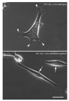
Morphology of normal 3T3 cells and Src knockout fibroblast (SYF) cells (c-Src, c-Yes, and Fyn knockout cells), as observed by phase-contrast microscopy. (a) The morphology of normal fibroblasts. (b) Fluorescence microscopy showing focal adhesions stained with anti-vinculin antibody. (c) When SYF cells were cultured on a glass substrate, they first showed a pancake-like morphology and then adopted a symmetrical spindle shape (arrowheads). (d) In this process, focal adhesions were formed at both ends of the cells, and a relatively small adhesive patch-like structure was observed at the center of the cells (arrows). (a,c) Phase-contrast microscopy. (b,d) Fluorescence microscopy showing focal adhesions stained with anti-vinculin antibody. Scale bars: (a,c), 100 μm; (b,d), 20 μm.
Figure 2
Figure 2
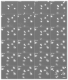
Normal fibroblasts cultured on a glass substrate in medium containing c-Src inhibitor (Src Inhibitor No. 5). Normal fibroblasts cultured in a medium containing 10 μM Src Inhibitor No. 5, a c-Src inhibitor, adhered to the glass substrate and then showed symmetrical spindle-like extension (arrowheads). The morphology was similar to that of SYF cells stretched on the glass substrate (see Figure 1c). Phase-contrast microscopy time-lapse images. The numbers at the top left indicate the number of minutes since the start of incubation. Scale bar, 100 μm. See also Video S1.
Figure 3
Figure 3
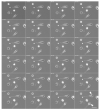
Morphological changes in normal fibroblasts after removal of c-Src inhibitor. After washout of the c-Src inhibitor with inhibitor-free culture medium, the elongated fibroblasts began to migrate in one direction, extending their pseudopods and eventually showing typical fibroblast morphology (arrowheads). The medium of the cells shown in Figure 2 was replaced with normal culture medium. The time intervals are shown in minutes at the top left corner of each time-lapse phase-contrast microscopy image. Scale bar, 100 μm. See also Video S2.
Figure 4
Figure 4
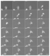
Normal fibroblasts expressing the dominant negative c-Src gene. The dominant negative c-Src gene (pUSEamp-dominant negative c-Src) was expressed in normal fibroblasts, and living cells were recorded under phase-contrast microscopy. The time intervals are shown in minutes at the top left corner of each time-lapse phase-contrast microscopy image. Scale bar, 100 μm. See also Video S3.
Figure 5
Figure 5
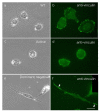
Normal fibroblasts expressing WT c-Src, constitutively active c-Src, and dominant negative c-Src genes. pUSEamp-WT c-Src (a,b), pUSEamp-constitutively active c-Src (c,d), and pUSEamp-dominant negative c-Src (e,f) were transfected into normal fibroblasts, and fixed cells were recorded under phase-contrast microscopy. Cells expressing the dominant negative c-Src gene were elongated at both poles in a symmetrical cone shape (e,f). (c) The cells expressing constitutively active c-Src show a pancake-like morphology. (a,c,e) Phase-contrast microscopy. (b,d,f) Fluorescence microscopy of cells stained with anti-vinculin antibody. Figure 1b shows normal fibroblasts stained with anti-vinculin antibody as a control. Scale bar, 20 μm.
Figure 6
Figure 6
SYF cells expressing eGFP-fused wild-type (WT) c-Src and eGFP-fused dominant negative c-Src genes. SYF cells expressing (a) eGFP-fused WT c-Src and (b) eGFP-fused dominant negative c-Src were observed by phase-contrast microscopy. Cells transfected with (a) eGFP-fused WT c-Src were elongated with pseudopodia (arrowheads) similar to normal fibroblasts, whereas (b) cells expressing eGFP-fused dominant negative c-Src showed an elongated symmetrical cone shape (arrows). Scale bar, 20 μm.
Figure 7
Figure 7
Elongation ratio of the vertical axis to the long axis.

Similar articles

Cited by

References

    1. Assémat E., Bazellières E., Pallesi-Pocachard E., Le Bivic A., Massey-Harroche D. Polarity complex proteins. Biochim. Biophys. Acta. 2008;1778:614–630. doi: 10.1016/j.bbamem.2007.08.029. - DOI - PubMed
    1. Welch M.D., Mullins R.D. Cellular control of actin nucleation. Annu. Rev. Cell Dev. Biol. 2002;18:247–288. doi: 10.1146/annurev.cellbio.18.040202.112133. - DOI - PubMed
    1. Pollard T.D., Borisy G.G. Cellular motility driven by assembly and disassembly of actin filaments. Cell. 2003;112:453–465. doi: 10.1016/S0092-8674(03)00120-X. - DOI - PubMed
    1. Katoh K., Kano Y., Fujiwara K. Isolation and in vitro contraction of stress fibers. Methods Enzymol. 2000;325:369–380. - PubMed
    1. Katoh K., Kano Y., Ookawara S. Rho-kinase dependent organization of stress fibers and focal adhesions in cultured fibroblasts. Genes Cells. 2007;12:623–638. doi: 10.1111/j.1365-2443.2007.01073.x. - DOI - PubMed

LinkOut - more resources